- ICC/IF
Supplier Data
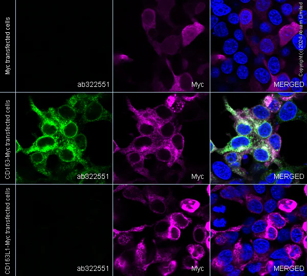
Immunocytochemistry/ Immunofluorescence - Anti-CD163 antibody [EPR29058-507] (AB322551)
Immunofluorescent analysis of 4% Paraformaldehyde-fixed, 0.1% TritonX-100 permeabilized 293T (human embryonic kidney epithelial cell) cells labelling CD163 with ab322551 at 1/5000 (0.1 ug/ml) dilution, followed by ab150081 Goat Anti-Rabbit IgG H&L (Alexa Fluor® 488) preadsorbed antibody at 1/1000 2ug/ml dilution (Green).
Confocal image showing cytoplasmic staining in 293T cells (shown in green) transfected with a human CD163 expression vector containing a myc-His-tag®, showing no staining in 293T cells transfected with a human CD163L or empty expression vector containing a myc-His-tag®. The counterstain was observed in magenta. Nuclear DNA was labelled with DAPI (shown in blue). Image was taken with a confocal microscope (Leica-Microsystems, TCS SP8).
ab223894 Anti-Myc tag mouse monoclonal antibody (Alexa Fluor® 594) was used to counterstain tubulin at 1/ab223894 100 5ug/ml dilution (Magenta). The Nuclear counterstain was DAPI (Blue).
Secondary antibody only control : Secondary antibody is ab150081 Goat Anti-Rabbit IgG H&L (Alexa Fluor® 488) preadsorbed at 1/1000 2ug/ml dilution.
- ICC/IF
Supplier Data
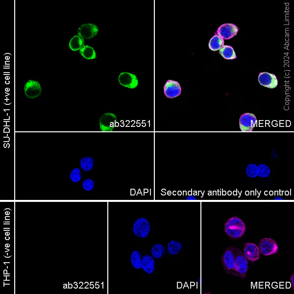
Immunocytochemistry/ Immunofluorescence - Anti-CD163 antibody [EPR29058-507] (AB322551)
Immunofluorescent analysis of 4% Paraformaldehyde-fixed, 0.1% TritonX-100 permeabilized SU-DHL-1(human histiocytic lymphoblast-like cell) cells labelling CD163 with ab322551 at 1/5000 (0.1 ug/ml) dilution, followed by ab150081 Goat Anti-Rabbit IgG H&L (Alexa Fluor® 488) preadsorbed antibody at 1/1000 2ug/ml dilution (Green).
Confocal image showing cytoplasmic staining in SU-DHL-1 cell line (shown in green). The counterstain was observed in magenta. Nuclear DNA was labelled with DAPI (shown in blue).
Negative control : THP-1(PMID : 10648003).
Image was taken with a confocal microscope (Leica-Microsystems, TCS SP8).
ab195889 Anti-alpha Tubulin mouse monoclonal antibody - Microtubule Marker (Alexa Fluor® 594) was used to counterstain tubulin at 1/ab195889 200 2.5ug/ml dilution (Magenta). The Nuclear counterstain was DAPI (Blue).
Secondary antibody only control : Secondary antibody is ab150081 Goat Anti-Rabbit IgG H&L (Alexa Fluor® 488) preadsorbed at 1/1000 2ug/ml dilution.
- ICC/IF
Supplier Data
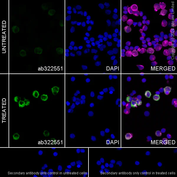
Immunocytochemistry/ Immunofluorescence - Anti-CD163 antibody [EPR29058-507] (AB322551)
Immunofluorescent analysis of 4% Paraformaldehyde-fixed, 0.1% TritonX-100 permeabilized Human PBMC (human primary peripheral blood mononuclear cell) cells labelling CD163 with ab322551 at 1/5000 (0.1 ug/ml) dilution, followed by ab150081 Goat Anti-Rabbit IgG H&L (Alexa Fluor® 488) preadsorbed antibody at 1/ 1000 2ug/ml dilution (Green).
Confocal image showing increased cytoplasmic staining in subsets of human PBMCs (shown in green) after treatment with 10 ng/mL IL-10 for 18 hours. The counterstain was observed in magenta. Nuclear DNA was labelled with DAPI (shown in blue). Image was taken with a confocal microscope (Leica-Microsystems, TCS SP8).
ab195889 Anti-alpha Tubulin mouse monoclonal antibody - Microtubule Marker (Alexa Fluor® 594) was used to counterstain tubulin at 1/ 200 2.5ug/ml dilution (Magenta). The Nuclear counterstain was DAPI (Blue).
Secondary antibody only control : Secondary antibody is ab150081 Goat Anti-Rabbit IgG H&L (Alexa Fluor® 488) preadsorbed at 1/ 1000 2ug/ml dilution.
- Flow Cyt
Supplier Data
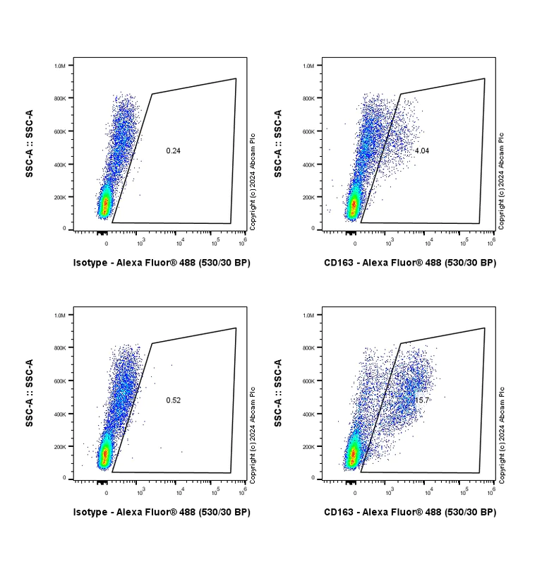
Flow Cytometry - Anti-CD163 antibody [EPR29058-507] (AB322551)
Flow cytometric analysis of Human PBMC(human peripheral blood mononuclear cell) untreated with 10 ng/mL IL-10 for 18 hours(up left and up right) / Human PBMC treated with 10 ng/mL IL-10 for 18 hours(down left and down right) cells labelling CD163 with ab322551 at 1/5000 dilution (0.01 ug)/down right compared with a Rabbit monoclonal IgG (ab172730) / Left isotype control and an unlabelled control (cells without incubation with primary antibody and secondary antibody).
Goat Anti-Rabbit IgG (Alexa Fluor® 488, ab150081) at 1/5000 dilution was used as the secondary antibody.
gated on viable cell.
보완 제품


Reactivity 정보
제품 세부 정보
Patented technology
Our RabMAb® technology is a patented hybridoma-based technology for making rabbit monoclonal antibodies. For details on our patents, please refer to RabMAb® patents.
What are the advantages of a recombinant monoclonal antibody?
This product is a recombinant monoclonal antibody, which offers several advantages including:
- - High batch-to-batch consistency and reproducibility
- - Improved sensitivity and specificity
- - Long-term security of supply
- - Animal-free batch production
For more information, read more on recombinant antibodies.
특성 및 보관 정보
제형
Purification 테크닉
보관 버퍼
배송 시 보관 조건
적절한 단기 보관 기간
적절한 단기 보관 조건
적절한 장기 보관 조건
분주 정보
보관 정보
제품 프로토콜
- Visit the General protocols
- Visit the Troubleshooting
타겟 정보
대체 명칭 보기
Product promise
Please note: All products are 'FOR RESEARCH USE ONLY. NOT FOR USE IN DIAGNOSTIC OR THERAPEUTIC PROCEDURES'.
For licensing inquiries, please contact partnerships@abcam.com