Anti-CD21 antibody [EPR27369-9]
- BOND RX™ Validated
- 20ul selling size
- Recombinant
- Advanced Validation
- RabMAb
- 각 태그의 뜻
Be the first to review this product! 리뷰 제출
|
(5 제품이 사용된 논문 )
- Flow Cyt
Supplier Data
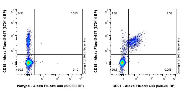
Flow Cytometry - Anti-CD21 antibody [EPR27369-9] (AB315160)
Flow cytometric analysis of Human PBMC (human peripheral blood mononuclear cell) cells labelling CD21 with ab315160 at 1/500 dilution (0.1 ug)/Right compared with a Rabbit monoclonal IgG (ab172730) / Left isotype control.
Goat Anti-Rabbit IgG (Alexa Fluor® 488, ab150081) at 1/5000 dilution was used as the secondary antibody.
Gated on viable cells. Cells are co-stained with CD19 conjugated to Alexa Fluor®647.
- Flow Cyt
Supplier Data
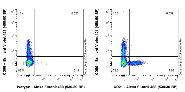
Flow Cytometry - Anti-CD21 antibody [EPR27369-9] (AB315160)
Flow cytometric analysis of Human PBMC (human peripheral blood mononuclear cell) cells labelling CD21 with ab315160 at 1/500 dilution (0.1 ug)/Right compared with a Rabbit monoclonal IgG (ab172730) / Left isotype control
Goat Anti-Rabbit IgG (Alexa Fluor® 488, ab150081) at 1/5000 dilution was used as the secondary antibody.
Gated on viable cells. Cells are co-stained with CD56 conjugated to Brilliant Violet 421.
- IHC-P
Supplier Data
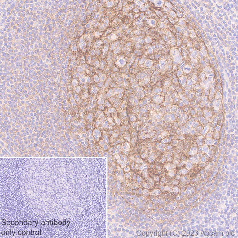
Immunohistochemistry (Formalin/PFA-fixed paraffin-embedded sections) - Anti-CD21 antibody [EPR27369-9] (AB315160)
Immunohistochemical analysis of paraffin-embedded Human tonsil tissue labeling CD21 with ab315160 at 1/50 (10.56 ug/ml) dilution, followed by a ready to use Rabbit specific IHC polymer detection kit HRP/DAB (ab209101).
Positive staining on germinal center of human tonsil. The section was incubated with ab315160 for 30 mins at room temperature. The immunostaining was performed on a Leica Biosystems BOND® RX instrument Counterstained with Hematoxylin.
Secondary antibody only control : Secondary antibody is a ready to use Rabbit specific IHC polymer detection kit HRP/DAB (ab209101).
Heat mediated antigen retrieval was performed with Tris-EDTA buffer (pH 9.0, Epitope Retrieval Solution2) for 20 mins
- IHC-P
Supplier Data
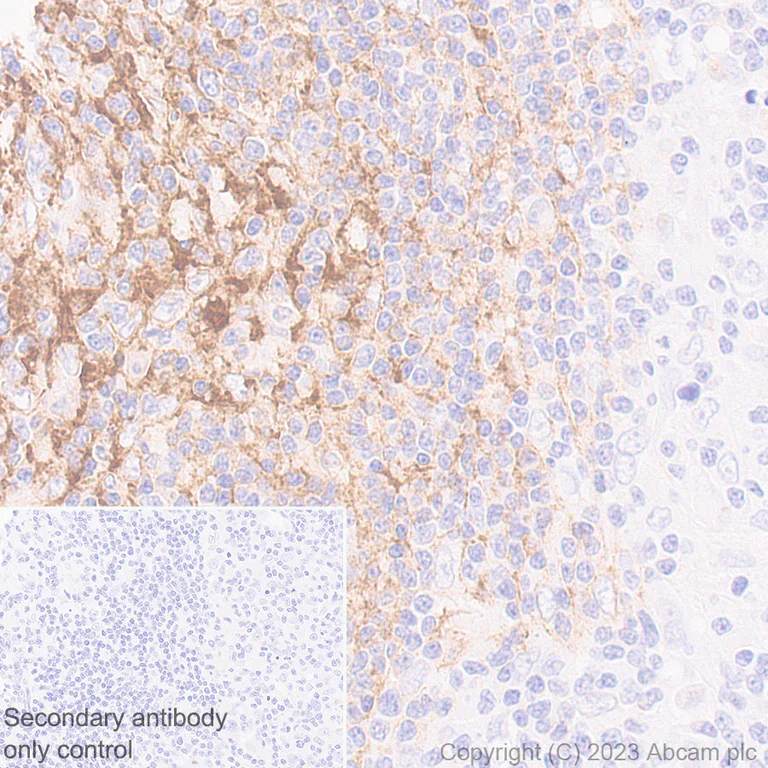
Immunohistochemistry (Formalin/PFA-fixed paraffin-embedded sections) - Anti-CD21 antibody [EPR27369-9] (AB315160)
Immunohistochemical analysis of paraffin-embedded Human hodgkin's lymphoma tissue labeling CD21 with ab315160 at 1/50 (10.56 ug/ml) dilution, followed by a ready to use Rabbit specific IHC polymer detection kit HRP/DAB (ab209101).
Positive staining on human hodgkin's lymphoma. The section was incubated with ab315160 for 30 mins at room temperature. The immunostaining was performed on a Leica Biosystems BOND® RX instrument Counterstained with Hematoxylin.
Secondary antibody only control : Secondary antibody is a ready to use Rabbit specific IHC polymer detection kit HRP/DAB (ab209101).
Heat mediated antigen retrieval was performed with Tris-EDTA buffer (pH 9.0, Epitope Retrieval Solution2) for 20 mins
- ICC/IF
Supplier Data
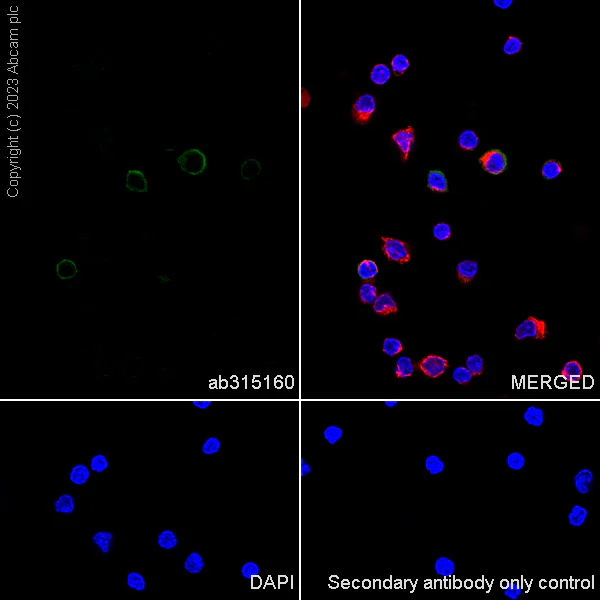
Immunocytochemistry/ Immunofluorescence - Anti-CD21 antibody [EPR27369-9] (AB315160)
Immunofluorescent analysis of 4% Paraformaldehyde-fixed, 0.1% Tween-20 permeabilized Human PBMC (human primary peripheral blood mononuclear cell) cells labelling CD21 with ab315160 at 1/50 (10.56 ug/ml) dilution, followed by ab150081 Goat Anti-Rabbit IgG H&L (Alexa Fluor® 488) preadsorbed antibody at 1/1000 (2 ug/ml) dilution (Green).
Confocal image showing membranous staining in subsets of human PBMCs. Image was taken with a confocal microscope(Leica-Microsystems, TCS SP8).
ab195889 Anti-alpha Tubulin mouse monoclonal antibody - Microtubule Marker (Alexa Fluor® 594) was used to counterstain tubulin at 1/200 (2.5 ug/ml) dilution (Red). The Nuclear counterstain was DAPI (Blue).
Secondary antibody only control : Secondary antibody is ab150081 Goat Anti-Rabbit IgG H&L (Alexa Fluor® 488) preadsorbed at 1/1000 (2 ug/ml) dilution.
- ICC/IF
Supplier Data
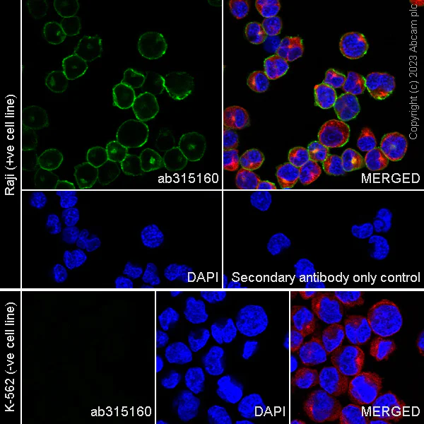
Immunocytochemistry/ Immunofluorescence - Anti-CD21 antibody [EPR27369-9] (AB315160)
Immunofluorescent analysis of 4% Paraformaldehyde-fixed, 0.1% Tween-20 permeabilized Raji (human Burkitt's lymphoma B lymphocyte) cells labelling CD21 with ab315160 at 1/50 (10.56 ug/ml) dilution, followed by ab150081 Goat Anti-Rabbit IgG H&L (Alexa Fluor® 488) preadsorbed antibody at 1/1000 (2 ug/ml) dilution (Green).
Confocal image showing membranous and cytoplasmic staining in Raji cell line. Negative control : K-562 (PMID : 6983911).Image was taken with a confocal microscope(Leica-Microsystems, TCS SP8).
ab195889 Anti-alpha Tubulin mouse monoclonal antibody - Microtubule Marker (Alexa Fluor® 594) was used to counterstain tubulin at 1/200 (2.5 ug/ml) dilution (Red). The Nuclear counterstain was DAPI (Blue).
Secondary antibody only control : Secondary antibody is ab150081 Goat Anti-Rabbit IgG H&L (Alexa Fluor® 488) preadsorbed at 1/1000 (2 ug/ml) dilution.
- IHC-P
Supplier Data
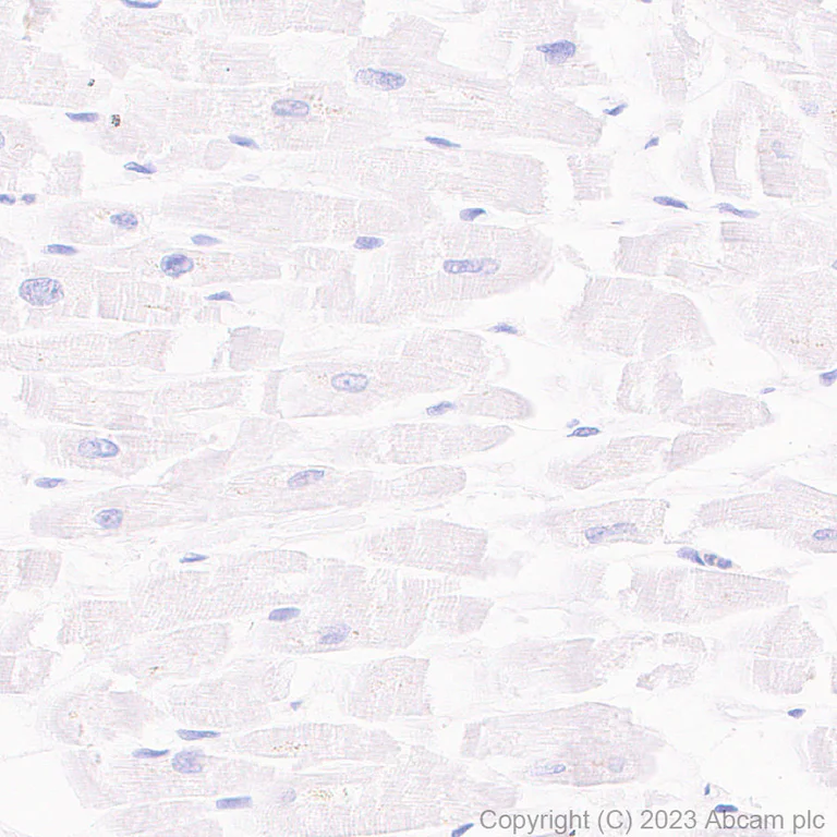
Immunohistochemistry (Formalin/PFA-fixed paraffin-embedded sections) - Anti-CD21 antibody [EPR27369-9] (AB315160)
Immunohistochemical analysis of paraffin-embedded Human cardiac muscle tissue labeling CD21 with ab315160 at 1/50 (10.56 ug/ml) dilution, followed by a ready to use Rabbit specific IHC polymer detection kit HRP/DAB (ab209101).
Negative control : no staining on human cardiac muscle. The section was incubated with ab315160 for 30 mins at room temperature. The immunostaining was performed on a Leica Biosystems BOND® RX instrument Counterstained with Hematoxylin.
Secondary antibody only control : Secondary antibody is a ready to use Rabbit specific IHC polymer detection kit HRP/DAB (ab209101).
Heat mediated antigen retrieval was performed with Tris-EDTA buffer (pH 9.0, Epitope Retrieval Solution2) for 20 mins
- Flow Cyt
Supplier Data
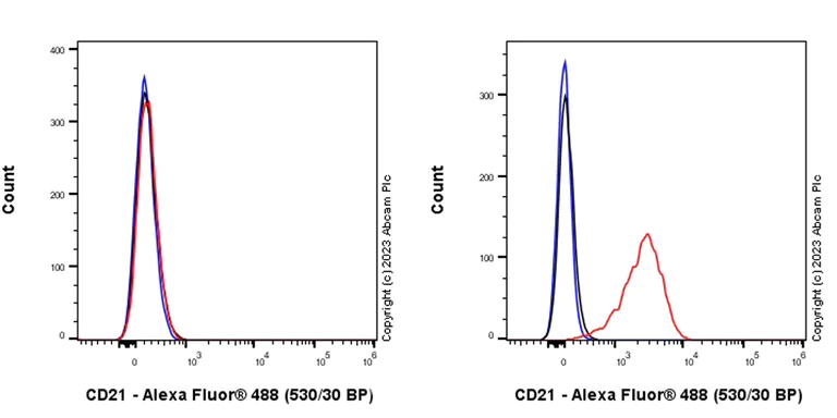
Flow Cytometry - Anti-CD21 antibody [EPR27369-9] (AB315160)
Flow cytometric analysis of K-562 (human chronic myelogenous leukemia lymphoblast, Left) / Raji (human Burkitt's lymphoma B lymphocyte, Right) cells labelling CD21 with ab315160 at 1/500 dilution (0.1 ug)/Red compared with a Rabbit monoclonal IgG (ab172730) (Black) isotype control and an unlabelled control (cells without incubation with primary antibody and secondary antibody) (Blue).
Goat Anti-Rabbit IgG (Alexa Fluor® 488, ab150081) at 1/5000 dilution was used as the secondary antibody.
Negative control : K-562. Gated on viable cells.
- mIHC
Supplier Data
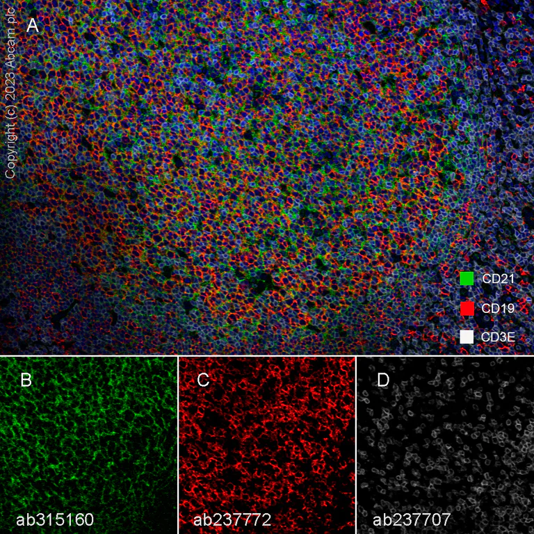
Multiplex immunohistochemistry - Anti-CD21 antibody [EPR27369-9] (AB315160)
Multiplex immunohistochemistry analysis of formalin/PFA-fixed paraffin-embedded human tonsil tissue staining CD21 with ab315160 at a 1 : 50 (10.56 ug/ml) dilution, ab237772 anti-CD19 used at 1 : 5000 (0.21 ug/ml) dilution and ab237707 anti-CD3E used at a 1 : 500 (1.01 ug/ml) dilution.
Panel A : merged staining of anti-CD21 (green; Opal™690), anti-CD3E (grey; Opal™520) and anti-CD19 (red; Opal™570) on human tonsil.
Panel B : anti-CD21 staining mature B lymphocytes and T-lymphocytes of human tonsil.
Panel C : anti-CD19 staining B lymphocytes of human tonsil.
Panel D : anti-CD3E staining T lymphocytes of human tonsil.
The section was incubated in three rounds of staining : in the order of ab315160, ab237772, and ab237707 for 30 mins at room temperature. Each round was followed by a separate fluorescent tyramide signal amplification system.
The immunostaining was performed on a Leica Biosystems BOND® RX instrument with an Opal™ 4-color kit. Image acquisition was performed with Leica SP8 confocal microscope.
Nuclear counter stain with DAPI.
Heat mediated antigen retrieval with Tris-EDTA buffer (pH 9.0, epitope retrieval solution2) for 20 mins.
- IP
Supplier Data
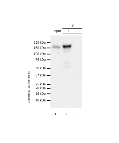
Immunoprecipitation - Anti-CD21 antibody [EPR27369-9] (AB315160)
CD21 was immunoprecipitated from 0.35 mg Raji (human Burkitt's lymphoma B lymphocyte) with ab315160 at 1/30 dilution (2ug in 0.35mg lysates). Western blot was performed on the immunoprecipitate using ab315160 at 1/1000 dilution. VeriBlot for IP secondary antibody(HRP)(ab131366) was used at 1/5000 dilution.
Lane 1 : Raji (human Burkitt's lymphoma B lymphocyte) whole cell lysate
Lane 2 : ab315160 IP in Raji (human Burkitt's lymphoma B lymphocyte)
Lane 3 : Rabbit monoclonal IgG (ab172730) instead of ab315160 in Raji whole cell lysate
Blocking and dilution buffer and concentration : 5% NFDM/TBST.
All lanes:
Immunoprecipitation - Anti-CD21 antibody [EPR27369-9] (ab315160) at 1/30 dilution
All lanes:
Raji (human Burkitt's lymphoma B lymphocyte) whole cell lysate
Secondary
All lanes:
Immunoprecipitation - VeriBlot for IP Detection Reagent (HRP) (<a href='/ko/products/reagents/veriblot-for-ip-detection-reagent-hrp-ab131366'>ab131366</a>) at 1/5000 dilution
false
Exposure time: 41s
- WB
Supplier Data
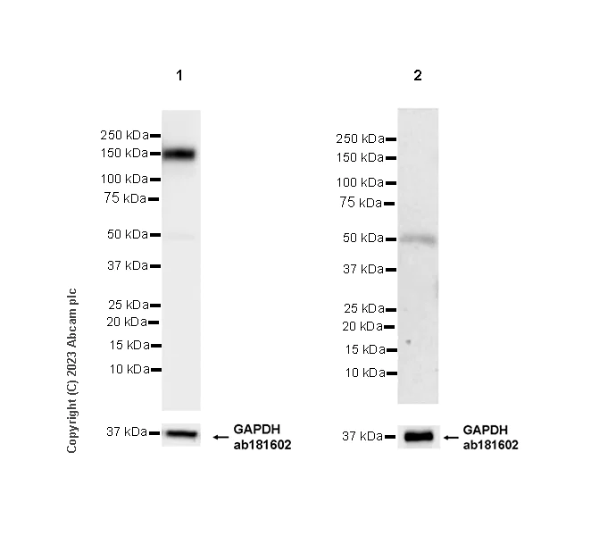
Western blot - Anti-CD21 antibody [EPR27369-9] (AB315160)
Blocking and diluting buffer and concentration : 5% NFDM/TBST.
Negative control : Human heart.
The identity of the lower MW bands at approximately 50kDa is unknown.
In Western blot, Anti-GAPDH antibody [EPR16891] - Loading Control (ab181602) staining at 1/200000 dilution.
Exposure time : Lane 1 : 15 seconds Lane 2 : 180 seconds
All lanes:
Western blot - Anti-CD21 antibody [EPR27369-9] (ab315160) at 1/1000 dilution
Lane 1:
Human tonsil tissue lysate at 20 µg
Lane 2:
Human heart tissue lysate at 20 µg
Secondary
All lanes:
Western blot - Goat Anti-Rabbit IgG H&L (HRP) (<a href='/ko/products/secondary-antibodies/goat-rabbit-igg-h-l-hrp-ab97051'>ab97051</a>) at 1/20000 dilution
Observed band size: 150 kDa,36 kDa
false
- WB
Supplier Data
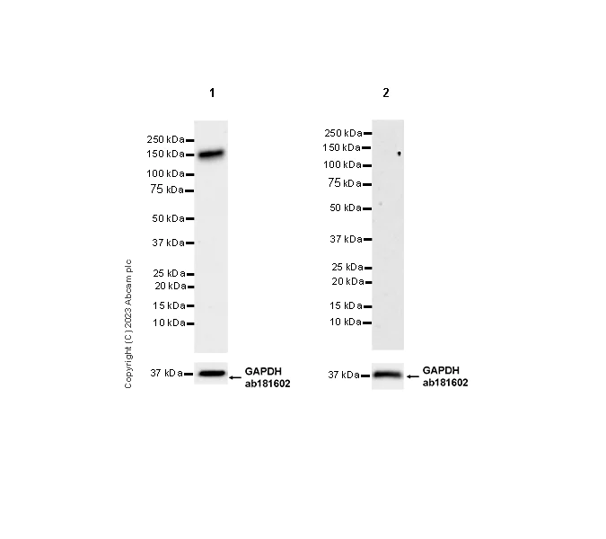
Western blot - Anti-CD21 antibody [EPR27369-9] (AB315160)
Blocking and diluting buffer and concentration : 5% NFDM/TBST.
Negative control : K-562 (PMID : 6983911).
In Western blot, Anti-GAPDH antibody [EPR16891] - Loading Control (ab181602) staining at 1/200000 dilution.
Exposure time : Lane 1 : 48 seconds Lane 2 : 180 seconds
All lanes:
Western blot - Anti-CD21 antibody [EPR27369-9] (ab315160) at 1/1000 dilution
Lane 1:
Raji (human Burkitt's lymphoma B lymphocyte) whole cell lysate at 20 µg
Lane 2:
K-562 (human chronic myelogenous leukemia lymphoblast) whole cell lysate at 20 µg
Secondary
All lanes:
Western blot - Goat Anti-Rabbit IgG H&L (HRP) (<a href='/ko/products/secondary-antibodies/goat-rabbit-igg-h-l-hrp-ab97051'>ab97051</a>) at 1/20000 dilution
Observed band size: 150 kDa,36 kDa
false
- WB
Supplier Data
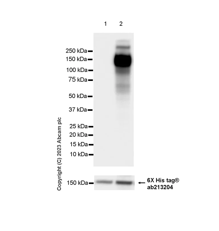
Western blot - Anti-CD21 antibody [EPR27369-9] (AB315160)
Blocking and diluting buffer and concentration : 5% NFDM/TBST.
In Western blot, Anti-6X His tag® antibody [EPR20547] - ChIP Grade (ab213204) staining at 1/5000 dilution.
All lanes:
Western blot - Anti-CD21 antibody [EPR27369-9] (ab315160) at 1/1000 dilution
Lane 1:
293T (human embryonic kidney epithelial cell) cells transfected with an empty vector containing a myc-His-tag®, whole cell lysate at 10 µg
Lane 2:
293T cells transfected with a human CD21 expression vector containing a myc-His-tag®, whole cell lysate at 10 µg
Secondary
All lanes:
Western blot - Goat Anti-Rabbit IgG H&L (HRP) (<a href='/ko/products/secondary-antibodies/goat-rabbit-igg-h-l-hrp-ab97051'>ab97051</a>) at 1/20000 dilution
Observed band size: 150 kDa
false
Exposure time: 3.25s
관련 conjugated 항체와 다양한 조성의 항체 (5)
-
Anti-CD21 antibody [EPR27369-9] - BSA and Azide free
-
519 Alexa Fluor® 488
Alexa Fluor® 488 Anti-CD21 antibody [EPR27369-9]
-
617 Alexa Fluor® 594
Alexa Fluor® 594 Anti-CD21 antibody [EPR27369-9]
-
665 Alexa Fluor® 647
Alexa Fluor® 647 Anti-CD21 antibody [EPR27369-9]
-
578 PE
PE Anti-CD21 antibody [EPR27369-9]
보완 제품
Primary Antibodies
AB133616
BOND RX™ Validated|RabMAb|Recombinant|Lab Essentials|20ul selling size
- 5
- 20
Primary Antibodies
AB16702
20ul selling size|BOND RX™ Validated|RabMAb|Recombinant
- 0
- 0
Primary Antibodies
AB16669
Anti-CD3 epsilon antibody [SP7]
BOND RX™ Validated|RabMAb|Advanced Validation|Recombinant|Lab Essentials|20ul selling size

- 4
- 80
Primary Antibodies
AB172610
Anti-Bcl6 antibody [EPR11410-43]
RabMAb|Recombinant|20ul selling size
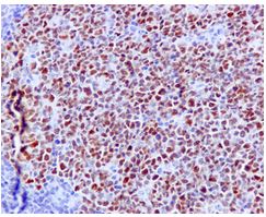
- 4
- 3
Reactivity 정보
제품 세부 정보
Patented technology
Our RabMAb® technology is a patented hybridoma-based technology for making rabbit monoclonal antibodies. For details on our patents, please refer to RabMAb® patents.
What are the advantages of a recombinant monoclonal antibody?
This product is a recombinant monoclonal antibody, which offers several advantages including:
- - High batch-to-batch consistency and reproducibility
- - Improved sensitivity and specificity
- - Long-term security of supply
- - Animal-free batch production
For more information, read more on recombinant antibodies.
특성 및 보관 정보
제형
Purification 테크닉
보관 버퍼
배송 시 보관 조건
적절한 단기 보관 기간
적절한 단기 보관 조건
적절한 장기 보관 조건
분주 정보
보관 정보
추가 정보
This supplementary information is collated from multiple sources and compiled automatically.
Biological function summary
Complement receptor type 2 is key in the immune system by facilitating B cell activation and proliferation. CD21 forms complexes with CD19 and CD81 increasing the sensitivity of B cells to antigens. This interaction enhances signal transduction enabling B lymphocytes to respond effectively to pathogens. The protein also plays a role in regulating immune tolerance and maintaining humoral immunity being important not only in immune defense but also in preventing autoimmunity.
Pathways
CD21 interacts with complement activation and immune response pathways. It connects with CD19 in a pivotal role where it further amplifies signaling in B lymphocytes. This interaction strengthens the downstream signaling of BCR (B cell receptor) important for antibody production. CD21 contributes to bridging the innate and adaptive immunological pathways by engaging components like C3d.
제품 프로토콜
- Visit the General protocols
- Visit the Troubleshooting
타겟 정보
대체 명칭 보기
제품이 사용된 논문 (5)
Recent publications for all applications. Explore the 전체 목록and refine your search
Nature communications 16:6960 PubMed40730843
2025
Applications
Unspecified application
Species
Unspecified reactive species
Journal of gastrointestinal oncology 16:386-403 PubMed40386587
2025
Applications
Unspecified application
Species
Unspecified reactive species
Oncology letters 28:468 PubMed39119236
2024
Applications
Unspecified application
Species
Unspecified reactive species
BMC cancer 24:758 PubMed38914961
2024
Applications
Unspecified application
Species
Unspecified reactive species
Frontiers in oncology 14:1383096 PubMed38846981
2024
Applications
Unspecified application
Species
Unspecified reactive species
Product promise
Please note: All products are 'FOR RESEARCH USE ONLY. NOT FOR USE IN DIAGNOSTIC OR THERAPEUTIC PROCEDURES'.
For licensing inquiries, please contact partnerships@abcam.com