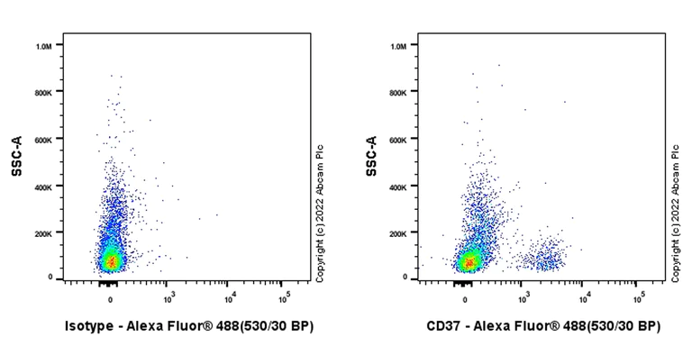
- Flow Cyt (Intra)
Supplier Data
Flow Cytometry (Intracellular) - Anti-CD37 antibody [EPR25397-149] (BSA and Azide free) (AB300412)
Flow cytometric analysis of Human peripheral blood mononuclear cell (PBMC) cells labelling CD37 with ab300400 at 1/500 dilution (0.1ug)/ Right compared with a Rabbit monoclonal IgG (ab172730) / Left isotype control and an unlabelled control (cells without incubation with primary antibody and secondary antibody) (Blue). Goat Anti-Rabbit IgG (Alexa Fluor® 488, ab150081) at 1/2000 dilution was used as the secondary antibody. Gated on viable cells.
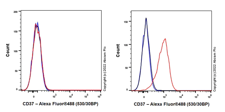
- Flow Cyt (Intra)
Supplier Data
Flow Cytometry (Intracellular) - Anti-CD37 antibody [EPR25397-149] (BSA and Azide free) (AB300412)
Flow cytometric analysis of MCF7 (human breast adenocarcinoma epithelial cell, Left) / Raji (human Burkitt's lymphoma B lymphocyte, Right) cells labelling CD37 with ab300400 at 1/500 dilution (0.1ug) (Red) compared with a Rabbit monoclonal IgG (ab172730) (Black) isotype control and an unlabelled control (cells without incubation with primary antibody and secondary antibody) (Blue). Goat Anti-Rabbit IgG (Alexa Fluor® 488, ab150081) at 1/2000 dilution was used as the secondary antibody. Gated on viable cells.Negative control : MCF7 (PMID : 33652767).
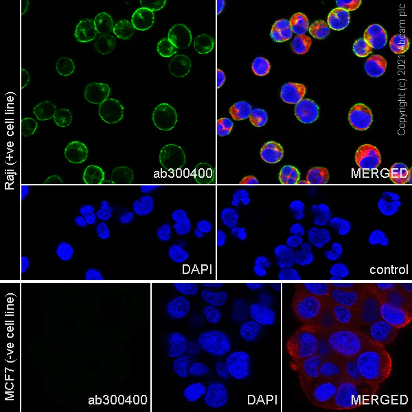
- ICC/IF
Supplier Data
Immunocytochemistry/ Immunofluorescence - Anti-CD37 antibody [EPR25397-149] (BSA and Azide free) (AB300412)
This data was developed using ab300400, the same antibody clone in a different buffer formulation.Immunofluorescent analysis of 100% methanol-fixed, 0.1% TritonX-100 permeabilized Raji (human Burkitt's lymphoma B lymphocyte) cells labelling CD37 with ab300400 at 1/500 (1.086 ug/ml) dilution, followed by ab150081 Goat Anti-Rabbit IgG H&L (Alexa Fluor® 488) preadsorbed antibody at 1/1000 2 ug/ml dilution (Green). Confocal image showing cytoplasmic and membranous staining in Raji cell line. Negative control : MCF7 (PMID : 33652767) is observed. ab195889 Anti-alpha Tubulin mouse monoclonal antibody - Microtubule Marker (Alexa Fluor® 594) was used to counterstain tubulin at 1/200 2.5ug/ml dilution (Red). The Nuclear counterstain was DAPI (Blue). Secondary antibody only control : Secondary antibody is ab150081 Goat Anti-Rabbit IgG H&L (Alexa Fluor® 488) preadsorbed at 1/1000 2 ug/ml dilution.
- IHC-P
Supplier Data
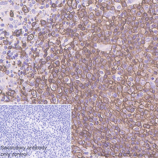
Immunohistochemistry (Formalin/PFA-fixed paraffin-embedded sections) - Anti-CD37 antibody [EPR25397-149] (BSA and Azide free) (AB300412)
This data was developed using ab300400, the same antibody clone in a different buffer formulation.Immunohistochemical analysis of paraffin-embedded Human Hodgkin's lymp tissue labeling CD37 with ab300400 at 1/500 (0.986 ug/ml) followed by a ready to use LeicaDS9800 (Bond Polymer Refine Detection) was used. Positive staining was found in human Hodgkin's lymphoma. The section was incubated with ab300400 for 30 mins at room temperature.The immunostaining was performed on a Leica Biosystems BOND® RX instrument Counterstained with Hematoxylin. Secondary antibody only control : Secondary antibody is a ready to use LeicaDS9800 (Bond Polymer Refine Detection) was used.Heat mediated antigen retrieval with Tris-EDTA buffer (pH 9.0, epitope retrieval solution2) for 20 mins
- IHC-P
Supplier Data
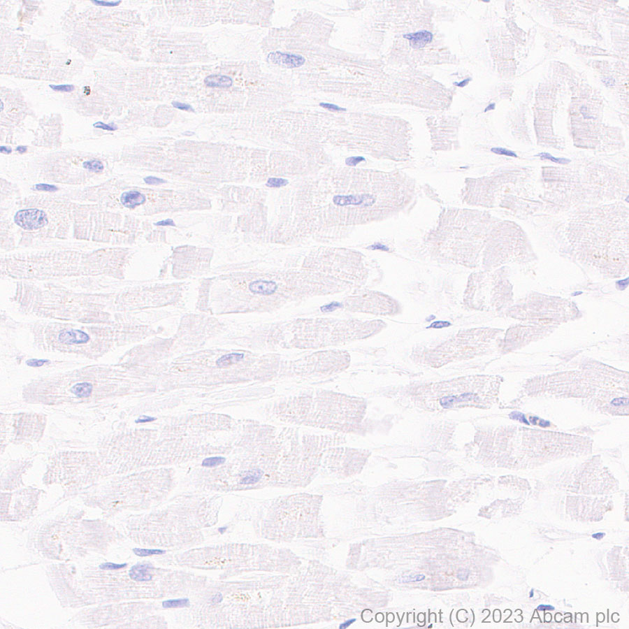
Immunohistochemistry (Formalin/PFA-fixed paraffin-embedded sections) - Anti-CD37 antibody [EPR25397-149] (BSA and Azide free) (AB300412)
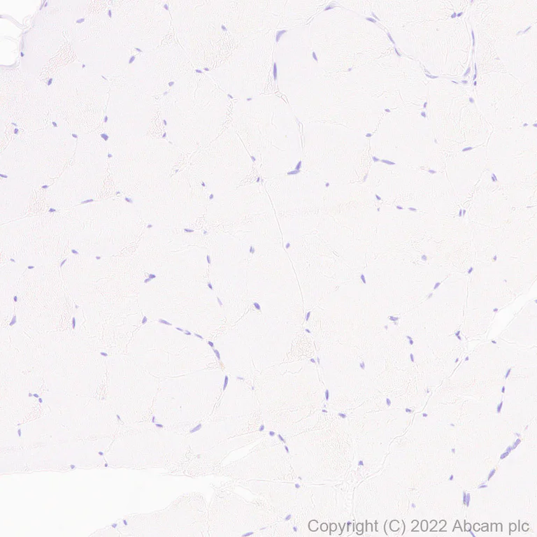
This data was developed using ab300400, the same antibody clone in a different buffer formulation.Immunohistochemical analysis of paraffin-embedded Human skeletal muscl tissue labeling CD37 with ab300400 at 1/500 (0.986 ug/ml) followed by a ready to use LeicaDS9800 (Bond Polymer Refine Detection) was used. Negative control : No staining was found on human skeletal muscle. The section was incubated with ab300400 for 30 mins at room temperature.The immunostaining was performed on a Leica Biosystems BOND® RX instrument Counterstained with Hematoxylin. Secondary antibody only control : Secondary antibody is a ready to use LeicaDS9800 (Bond Polymer Refine Detection) was used.Heat mediated antigen retrieval with Tris-EDTA buffer (pH 9.0, epitope retrieval solution2) for 20 mins
- IHC-P
Supplier Data
Immunohistochemistry (Formalin/PFA-fixed paraffin-embedded sections) - Anti-CD37 antibody [EPR25397-149] (BSA and Azide free) (AB300412)
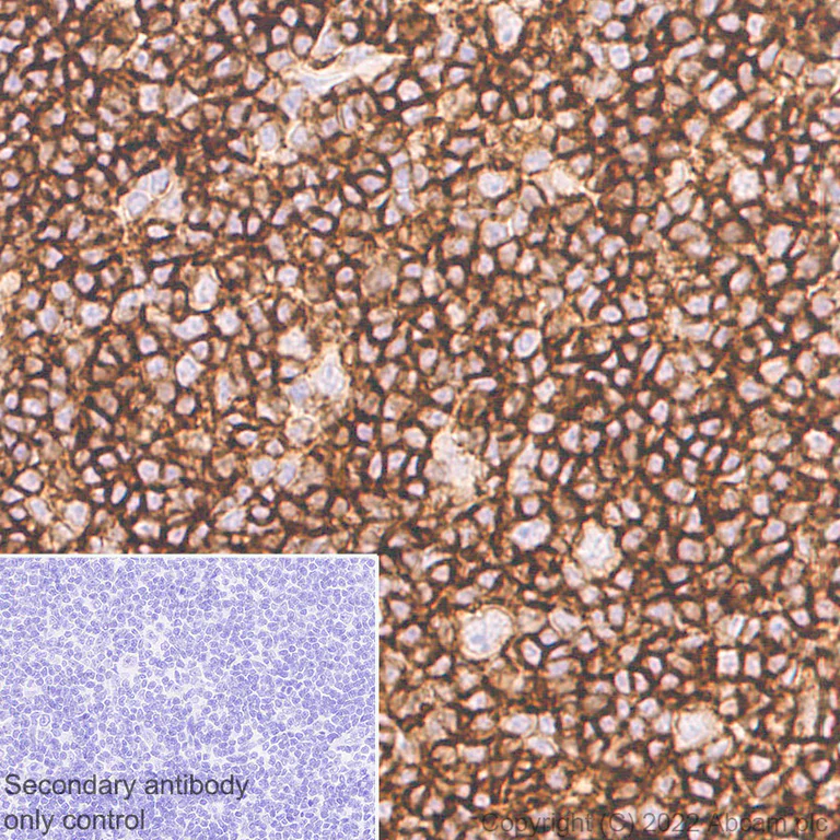
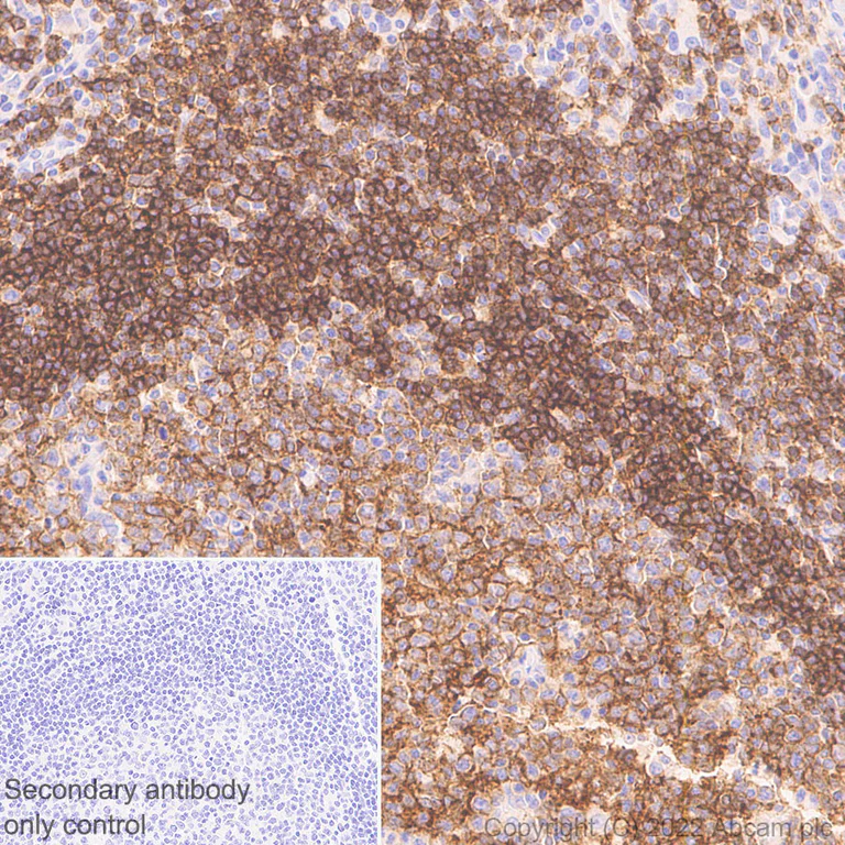
This data was developed using ab300400, the same antibody clone in a different buffer formulation.Immunohistochemical analysis of paraffin-embedded Human tonsil tissue labeling CD37 with ab300400 at 1/500 (0.986 ug/ml) followed by a ready to use LeicaDS9800 (Bond Polymer Refine Detection) was used. Positive staining was found in human tonsil (PMID : 25934707). The section was incubated with ab300400 for 30 mins at room temperature.The immunostaining was performed on a Leica Biosystems BOND® RX instrument Counterstained with Hematoxylin. Secondary antibody only control : Secondary antibody is a ready to use LeicaDS9800 (Bond Polymer Refine Detection) was used.Heat mediated antigen retrieval with Tris-EDTA buffer (pH 9.0, epitope retrieval solution2) for 20 mins
- IHC-P
Supplier Data
Immunohistochemistry (Formalin/PFA-fixed paraffin-embedded sections) - Anti-CD37 antibody [EPR25397-149] (BSA and Azide free) (AB300412)
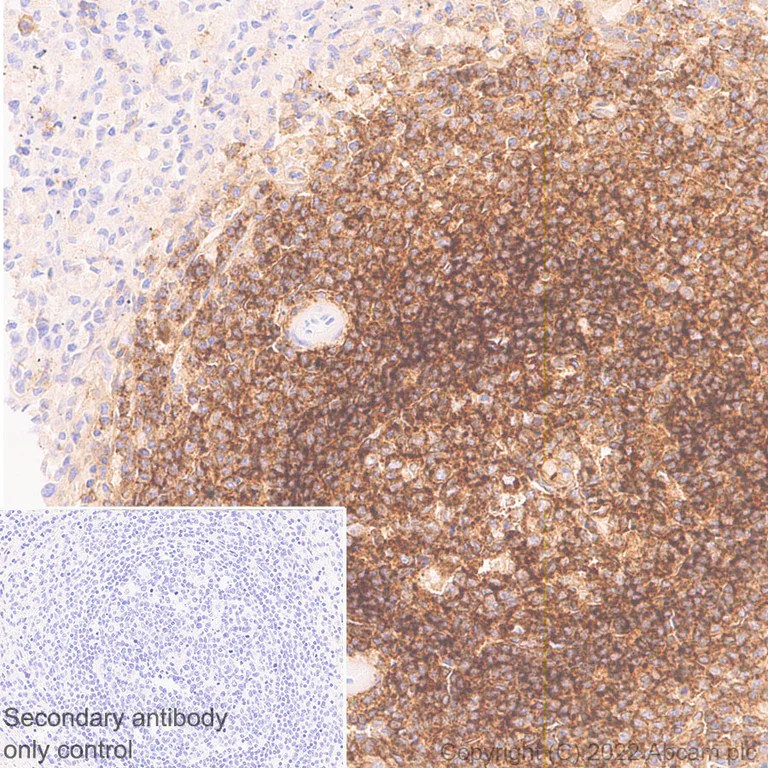
This data was developed using ab300400, the same antibody clone in a different buffer formulation.Immunohistochemical analysis of paraffin-embedded Human spleen tissue labeling CD37 with ab300400 at 1/500 (0.986 ug/ml) followed by a ready to use LeicaDS9800 (Bond Polymer Refine Detection) was used. Positive staining was found in human spleen (PMID : 25934707). The section was incubated with ab300400 for 30 mins at room temperature.The immunostaining was performed on a Leica Biosystems BOND® RX instrument Counterstained with Hematoxylin. Secondary antibody only control : Secondary antibody is a ready to use LeicaDS9800 (Bond Polymer Refine Detection) was used.Heat mediated antigen retrieval with Tris-EDTA buffer (pH 9.0, epitope retrieval solution2) for 20 mins
- Flow Cyt (Intra)
Supplier Data
Flow Cytometry (Intracellular) - Anti-CD37 antibody [EPR25397-149] (BSA and Azide free) (AB300412)
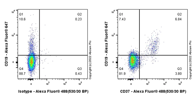
Flow cytometric analysis of Human peripheral blood mononuclear cell (PBMC) cells labelling CD37 with ab300400 at 1/500 dilution (0.1ug)/ Right compared with a Rabbit monoclonal IgG (ab172730) / Left isotype control. Goat Anti-Rabbit IgG (Alexa Fluor® 488, ab150081) at 1/2000 dilution was used as the secondary antibody. Gated on viable cells. Cells were stained with rabbit IgG or ab300400. Then stained with anti-CD19 conjugated to Alexa Fluor® 647.
- IP
Supplier Data
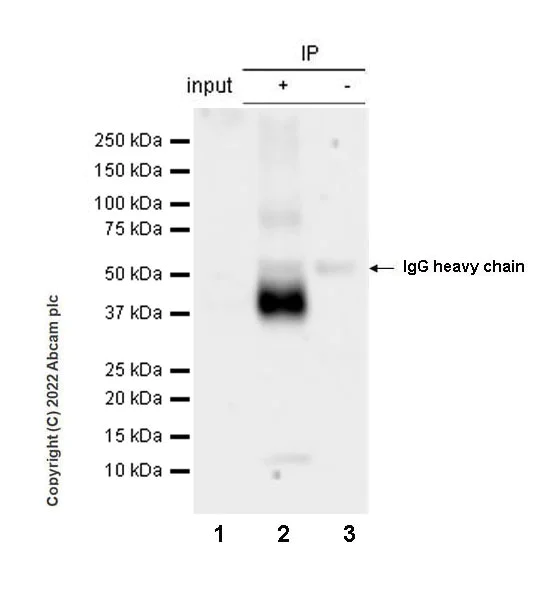
Immunoprecipitation - Anti-CD37 antibody [EPR25397-149] (BSA and Azide free) (AB300412)
This data was developed using ab300400, the same antibody clone in a different buffer formulation. CD37 was immunoprecipitated from 0.35 mg Human tonsil tissue lysate 10 ug with ab300400 at 1/30 dilution (2ug in 0.35mg lysates). Western blot was performed on the immunoprecipitate using ab300400 at 1/1000 dilution. VeriBlot for IP secondary antibody (HRP) (ab131366) was used at 1/5000 dilution.
Lane 1 : Human tonsil tissue lysate 10 ug
Lane 2 : ab300400 IP in Human tonsil tissue lysate
Lane 3 : Rabbit monoclonal IgG (ab172730) instead of ab300400 in Human tonsil tissue lysate
Blocking and dilution buffer and concentration : 5% NFDM/TBST.
Exposure time : 15 seconds
All lanes:
Immunoprecipitation - Anti-CD37 antibody [EPR25397-149] (<a href='/ko/products/primary-antibodies/cd37-antibody-epr25397-149-ab300400'>ab300400</a>) at 1/1000 dilution
All lanes:
Human tonsil tissue lysate at 10 µg
Secondary
All lanes:
Immunoprecipitation - VeriBlot for IP Detection Reagent (HRP) (<a href='/ko/products/reagents/veriblot-for-ip-detection-reagent-hrp-ab131366'>ab131366</a>) at 1/5000 dilution
false
Exposure time: 15s
- WB
Supplier Data
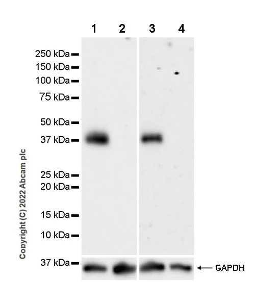
Western blot - Anti-CD37 antibody [EPR25397-149] (BSA and Azide free) (AB300412)
This data was developed using ab300400, the same antibody clone in a different buffer formulation. Blocking Buffer and concentration : 5% NFDM/TBST
All lanes:
Western blot - Anti-CD37 antibody [EPR25397-149] (<a href='/ko/products/primary-antibodies/cd37-antibody-epr25397-149-ab300400'>ab300400</a>) at 1/1000 dilution
Lane 1:
Human tonsil tissue lysate
Lane 2:
Human skeletal muscle tissue lysate
Lane 3:
Raji (human Burkitts lymphoma B lymphocyte), whole cell lysate
Lane 4:
MCF7 (human breast adenocarcinoma epithelial cell), whole cell lysate
Secondary
All lanes:
Western blot - Goat Anti-Rabbit IgG H&L (HRP) (<a href='/ko/products/secondary-antibodies/goat-rabbit-igg-h-l-hrp-ab97051'>ab97051</a>) at 1/100000 dilution
Observed band size: 40 kDa
false
관련 conjugated 항체와 다양한 조성의 항체 (1)
-
Anti-CD37 antibody [EPR25397-149]
보완 제품
Primary Antibodies
AB64088
BOND RX™ Validated|20ul selling size|RabMAb|Advanced Validation|Recombinant|KO Validated
- 5
- 13
Primary Antibodies
AB315160
Anti-CD21 antibody [EPR27369-9]
BOND RX™ Validated|20ul selling size|Recombinant|Advanced Validation|RabMAb
- 0
- 0
Primary Antibodies
AB16669
Anti-CD3 epsilon antibody [SP7]
BOND RX™ Validated|RabMAb|Advanced Validation|Recombinant|Lab Essentials|20ul selling size

- 4
- 80
Primary Antibodies
AB134147
BOND RX™ Validated|RabMAb|Recombinant|20ul selling size

- 2
- 1
Reactivity 정보
제품 세부 정보
Compatibility
This product is compatible with the Maxpar® Antibody Labeling Kit from Fluidigm, without the need for antibody preparation. Maxpar® is a trademark of Fluidigm Canada Inc.
Conjugation ready
Our carrier-free antibodies are typically supplied in a PBS-only formulation, purified and free of BSA, sodium azide and glycerol. This conjugation-ready format is designed for use with fluorochromes, metal isotopes, oligonucleotides, and enzymes, which makes them ideal for antibody labelling, functional and cell-based assays, flow-based assays (e.g. mass cytometry) and Multiplex Imaging applications.
Use our conjugation kits for antibody conjugates that are ready-to-use in as little as 20 minutes with 1 minute hands-on-time and 100% antibody recovery: available for fluorescent dyes, HRP, biotin and gold.
Patented technology
Our RabMAb® technology is a patented hybridoma-based technology for making rabbit monoclonal antibodies. For details on our patents, please refer to RabMAb® patents.
What are the advantages of a recombinant monoclonal antibody?
This product is a recombinant monoclonal antibody, which offers several advantages including:
- - High batch-to-batch consistency and reproducibility
- - Improved sensitivity and specificity
- - Long-term security of supply
- - Animal-free batch production
For more information, read more on recombinant antibodies.
특성 및 보관 정보
제형
Purification 테크닉
보관 버퍼
배송 시 보관 조건
적절한 단기 보관 조건
적절한 장기 보관 조건
제품 프로토콜
- Visit the General protocols
- Visit the Troubleshooting
타겟 정보
대체 명칭 보기
Product promise
Please note: All products are 'FOR RESEARCH USE ONLY. NOT FOR USE IN DIAGNOSTIC OR THERAPEUTIC PROCEDURES'.
For licensing inquiries, please contact partnerships@abcam.com