Anti-CD8 alpha antibody [EPR22483-288]
- BOND RX™ Validated
- 20ul selling size
- Recombinant
- Advanced Validation
- RabMAb
- 각 태그의 뜻
5
(1 리뷰)
|
(31 제품이 사용된 논문 )
- Multiplex IHC validated on the Leica BOND® MAX using Opal reagents
- Biophysical QC for unrivalled batch-batch consistency

- mIHC
Lab
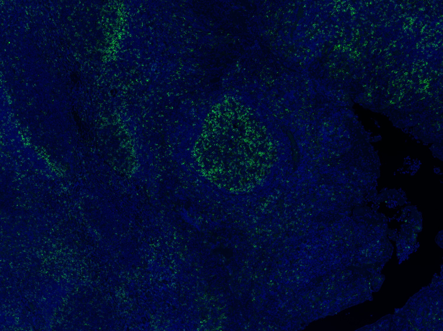
Multiplex immunohistochemistry - Anti-CD8 alpha antibody [EPR22483-288] (AB245118)
Multiplex immunohistochemistry analysis of formalin/PFA-fixed paraffin-embedded Human tonsil staining of PVRIG/CD112R with ab322348 at a 1/100 (5.16 µg/ml) dilution, CD19 with ab320735 at 1/2000 (0.255 µg/ml) dilution and CD8A with ab245118 at 1/1000 ( 0.375 µg/ml) dilution.
Panel A : merged staining of anti-PVRIG/CD112R (green; Opal™520), anti-CD8A (magenta; Opal™570) and anti-CD19 (yellow; Opal™690) on human tonsil.
Panel B : anti-PVRIG/CD112R staining memory and effector CD8+ T lymphocytes in human tonsil.
Panel C : anti-CD8A staining CD8+ T lymphocytes in human tonsil.
Panel D : anti-CD19 staining B lymphocytes in human tonsil.
Nuclear DNA was labeled with DAPI (shown in blue). ).
The section was incubated in three rounds of staining : in the order of ab322348, ab245118 and ab320735 for 30 mins at room temperature. Each round was followed by a separate fluorescent tyramide signal amplification system.
Heat mediated antigen retrieval with Tris-EDTA buffer (pH 9.0 epitope retrieval solution2) for 20 mins.
- mIHC
Lab
Multiplex immunohistochemistry - Anti-CD8 alpha antibody [EPR22483-288] (AB245118)
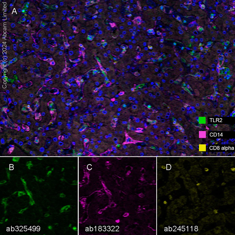
Multiplex immunohistochemistry analysis of formalin/PFA-fixed paraffin-embedded human liver tissue staining TLR2 with ab325499 at a 1 : 100 (5.0 µg/ml) dilution, ab183322 anti-CD14 used at 1 : 100 (1.22 µg/ml) dilution and ab245118 anti-CD8 alpha used at a 1 : 500 (0.75 µg/ml) dilution.
Panel A : anti-TLR2 (green; Opal™520), anti-CD14 (magenta; Opal™570) and anti-CD8 alpha (yellow; Opal™690) on human liver.
Panel B : anti-TLPR2 staining immune cells in human liver.
Panel C : anti-CD14 staining macrophages in human liver.
Panel D : anti-CD8 alpha staining T lymphocytes in human liver.
Nuclear DNA was labeled with DAPI (shown in blue).
The section was incubated in three rounds of staining : in the order of ab325499, ab183322 and ab245118 for 30 mins at room temperature. Each round was followed by a separate fluorescent tyramide signal amplification system.
The immunostaining was performed on a Leica Biosystems BOND® RX instrument with an Opal™ 4-color kit. Image acquisition was performed with Leica SP8 confocal microscope.
Nuclear counter stain with DAPI.
Heat mediated antigen retrieval was performed with Tris-EDTA buffer (pH 9.0, Epitope Retrieval Solution2) for 20 mins.
- mIHC
Supplier Data
Multiplex immunohistochemistry - Anti-CD8 alpha antibody [EPR22483-288] (AB245118)
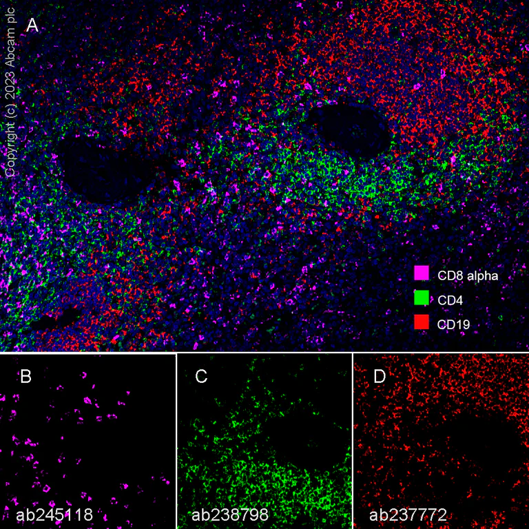
Multiplex immunohistochemistry analysis of formalin/PFA-fixed paraffin-embedded Human spleen tissue labeling CD8 alpha with ab245118 at 1/500 dilution, CD4 with ab238798 at 1/500, and CD19 with ab237772 at 1/5000 dilution.
Panel A : merged staining of anti-CD8 alpha (magenta; Opal™690), anti-CD4 (green; Opal™520) and anti-CD19 (red; Opal™570) on human spleen.
Panel B : anti-CD8 alpha stained on cytotoxic T cells.
Panel C : anti-CD4 stained on T helper cells.
Panel D : anti-CD19 stained on B cells.
Sections were treated using heat mediated antigen retrieval with Tris-EDTA buffer (pH 9.0, epitope retrieval solution2) for 20 mins before antibody incubation. The section was incubated in three rounds of staining : in the order of ab245118 for 30 mins, then ab238798 and ab237772 for 10 mins at room temperature. Each round was followed by a separate fluorescent tyramide signal amplification system.
DAPI was used as a nuclear counterstain.
The immunostaining was performed on a Leica Biosystems BOND® RX instrument with an Opal™ 4-color kit. Image acquisition was performed with Leica SP8 confocal microscope.
- mIHC
Supplier Data
Multiplex immunohistochemistry - Anti-CD8 alpha antibody [EPR22483-288] (AB245118)
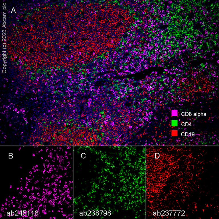
Multiplex immunohistochemistry analysis of formalin/PFA-fixed paraffin-embedded Human tonsil tissue labeling CD8 alpha with ab245118 at 1/500 dilution, CD4 with ab238798 at 1/500, and CD19 with ab237772 at 1/5000 dilution.
Panel A : merged staining of anti-CD8 alpha (magenta; Opal™690), anti-CD4 (green; Opal™520) and anti-CD19 (red; Opal™570) on human tonsil.
Panel B : anti-CD8 alpha stained on cytotoxic T cells.
Panel C : anti-CD4 stained on T helper cells.
Panel D : anti-CD19 stained on B cells.
Sections were treated using heat mediated antigen retrieval with Tris-EDTA buffer (pH 9.0, epitope retrieval solution2) for 20 mins before antibody incubation. The section was incubated in three rounds of staining : in the order of ab245118 for 30 mins, then ab238798 and ab237772 for 10 mins at room temperature. Each round was followed by a separate fluorescent tyramide signal amplification system.
DAPI was used as a nuclear counterstain.
The immunostaining was performed on a Leica Biosystems BOND® RX instrument with an Opal™ 4-color kit. Image acquisition was performed with Leica SP8 confocal microscope.
- Flow Cyt
Unknown
Flow Cytometry - Anti-CD8 alpha antibody [EPR22483-288] (AB245118)
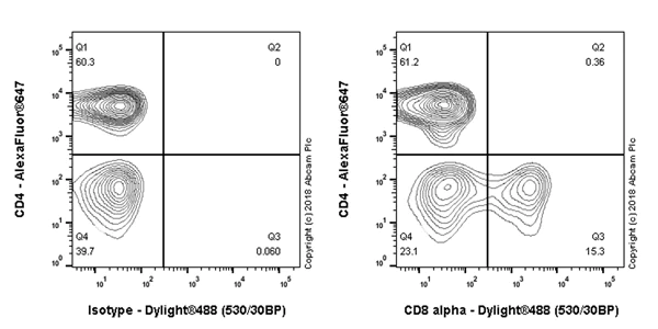
Flow cytometric analysis of human peripheral blood mononuclear cells (PBMC) labeling CD8 alpha with ab245118 at 1/400 (right panel) compared with a Rabbit monoclonal IgG (ab172730) (left panel). Goat anti rabbit IgG (Dylight® 488, ab98462) at 1/2000 dilution was used as the secondary antibody.
Cells were stained with rabbit IgG (Left) or anti-CD8 alpha RabMab (Right). Then stained with Alexa Fluor® 647-conjugated anti-CD4. The expressions of CD4 and CD8 alpha are mutually exclusive.
Gated on viable lymphocytes.
- IHC-P
Unknown
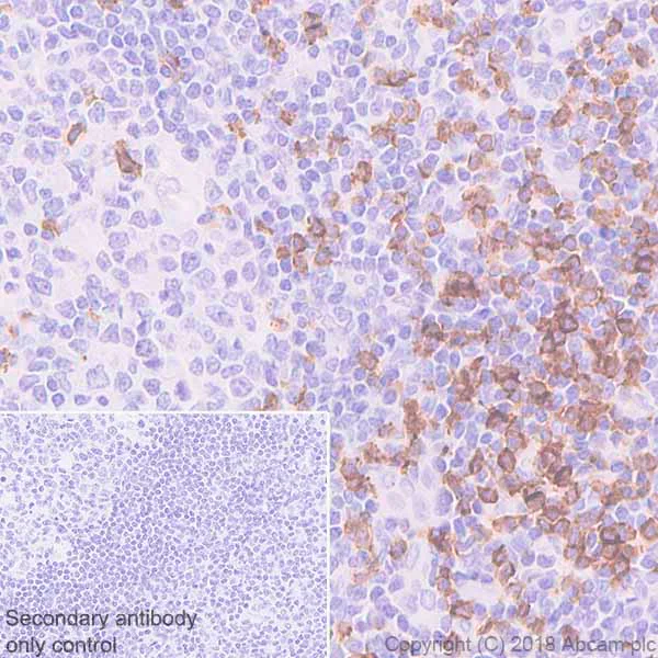
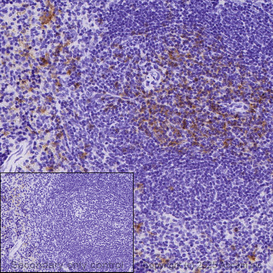
Immunohistochemistry (Formalin/PFA-fixed paraffin-embedded sections) - Anti-CD8 alpha antibody [EPR22483-288] (AB245118)
Immunohistochemical analysis of paraffin-embedded human tonsil tissue labeling CD8 alpha with ab245118 at 1/1000 dilution, followed by a ready to use Goat Anti-Rabbit IgG H&L (HRP). Positive staining on the T cell zone of human tonsil (PMID : 16504163) is observed. Counterstained with hematoxylin.
Secondary antibody only control : Used PBS instead of primary antibody, secondary antibody is a ready to use Goat Anti-Rabbit IgG H&L (HRP).
Perform heat mediated antigen retrieval using ab93684 (Tris/EDTA buffer, pH 9.0).
- IHC-P
Unknown
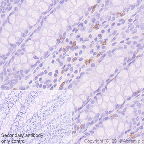
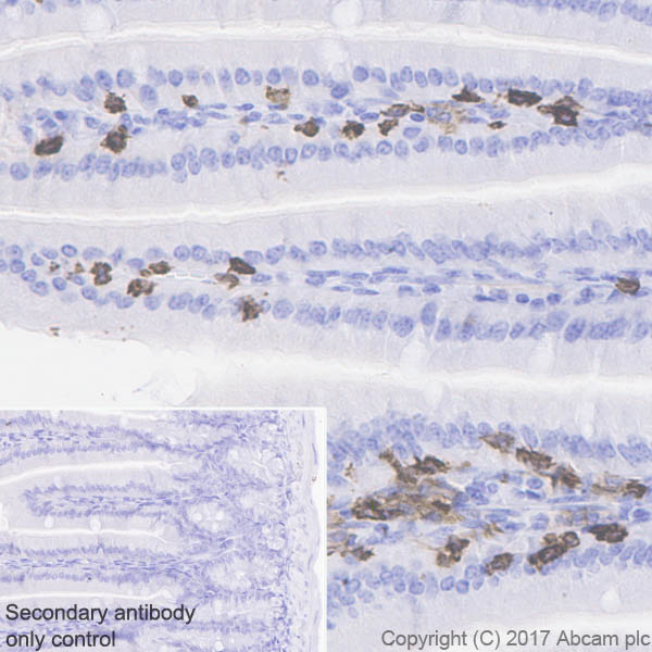
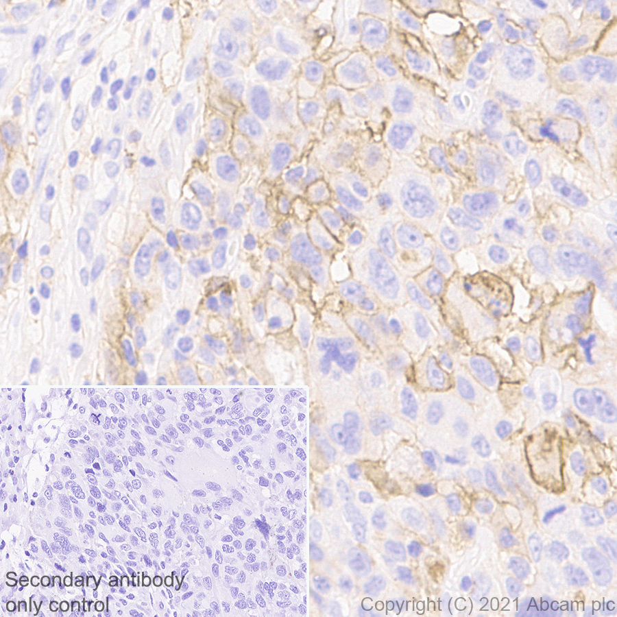
Immunohistochemistry (Formalin/PFA-fixed paraffin-embedded sections) - Anti-CD8 alpha antibody [EPR22483-288] (AB245118)
Immunohistochemical analysis of paraffin-embedded human colon tissue labeling CD8 alpha with ab245118 at 1/1000 dilution, followed by a ready to use Goat Anti-Rabbit IgG H&L (HRP). Positive staining on lymphocytes of human colon (PMID : 26310568) is observed. Counterstained with hematoxylin.
Secondary antibody only control : Used PBS instead of primary antibody, secondary antibody is a ready to use Goat Anti-Rabbit IgG H&L (HRP).
Perform heat mediated antigen retrieval using ab93684 (Tris/EDTA buffer, pH 9.0).
- mIHC
Lab
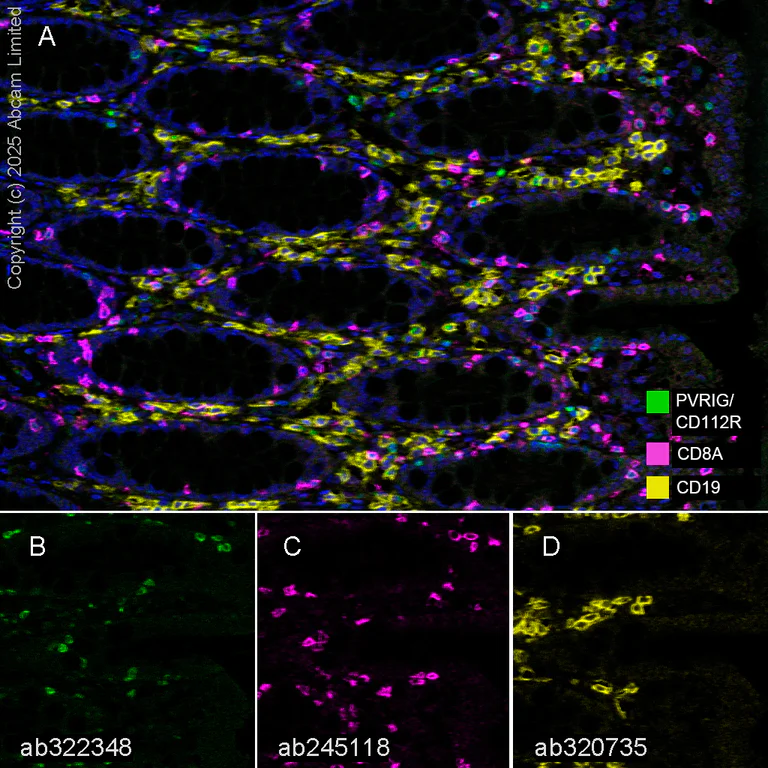
Multiplex immunohistochemistry - Anti-CD8 alpha antibody [EPR22483-288] (AB245118)
Multiplex immunohistochemistry analysis of formalin/PFA-fixed paraffin-embedded Human colon staining of PVRIG/CD112R with ab322348 at a 1/100 (5.16 µg/ml) dilution, CD19 with ab320735 at 1/2000 (0.255 µg/ml) dilution and CD8A with ab245118 at 1/1000 ( 0.375 µg/ml) dilution.
Panel A : merged staining of anti-PVRIG/CD112R (green; Opal™520), anti-CD8A (magenta; Opal™570) and anti-CD19 (yellow; Opal™690) on Human colon.
Panel B : anti-PVRIG/CD112R staining memory and effector CD8+ T lymphocytes in Human colon.
Panel C : anti-CD8A staining CD8+ T lymphocytes in Human colon.
Panel D : anti-CD19 staining B lymphocytes in Human colon.
Nuclear DNA was labelled with DAPI (shown in blue). ).
The section was incubated in three rounds of staining : in the order of ab322348, ab245118 and ab320735 for 30 mins at room temperature. Each round was followed by a separate fluorescent tyramide signal amplification system.
Heat mediated antigen retrieval with Tris-EDTA buffer (pH 9.0 epitope retrieval solution2) for 20 mins.
- mIHC
Lab
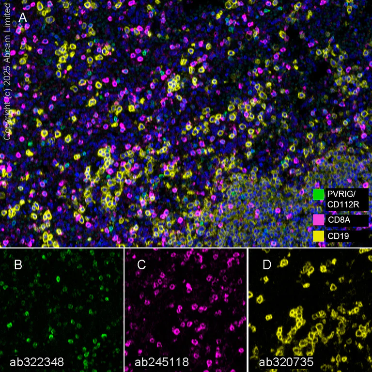
Multiplex immunohistochemistry - Anti-CD8 alpha antibody [EPR22483-288] (AB245118)
Multiplex immunohistochemistry analysis of formalin/PFA-fixed paraffin-embedded Human spleen staining of PVRIG/CD112R with ab322348 at a 1/100 (5.16 µg/ml) dilution, CD19 with ab320735 at 1/2000 (0.255 µg/ml) dilution and CD8A with ab245118 at 1/1000 ( 0.375 µg/ml) dilution.
Panel A : merged staining of anti-PVRIG/CD112R (green; Opal™520), anti-CD8A (magenta; Opal™570) and anti-CD19 (yellow; Opal™690) on human spleen.
Panel B : anti-PVRIG/CD112R staining memory and effector CD8+ T lymphocytes in human spleen.
Panel C : anti-CD8A staining CD8+ T lymphocytes in human spleen.
Panel D : anti-CD19 staining B lymphocytes in human spleen.
Nuclear DNA was labelled with DAPI (shown in blue). ).
The section was incubated in three rounds of staining : in the order of ab322348, ab245118 and ab320735 for 30 mins at room temperature. Each round was followed by a separate fluorescent tyramide signal amplification system.
Heat mediated antigen retrieval with Tris-EDTA buffer (pH 9.0 epitope retrieval solution2) for 20 mins.
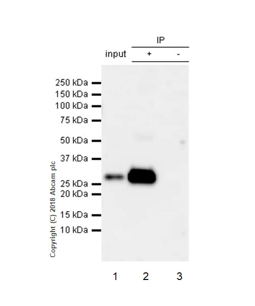
- IP
Unknown
Immunoprecipitation - Anti-CD8 alpha antibody [EPR22483-288] (AB245118)
CD8 alpha was immunoprecipitated from 0.35 mg human thymus lysate with ab245118 at 1/30 dilution. Western blot was performed from the immunoprecipitate using ab245118 at 1/500 dilution. VeriBlot for IP Detection Reagent (HRP) (ab131366), was used for detection at 1/1000 dilution.
Lane 1 : Human thymus lysate 10 μg (Input).
Lane 2 : ab245118 IP in human thymus lysate.
Lane 3 : Rabbit monoclonal IgG (ab172730) instead of ab245118 in human thymus lysate.
Blocking/Dilution buffer : 5% NFDM/TBST.
Exposure time : 30 seconds.
All lanes:
Immunoprecipitation - Anti-CD8 alpha antibody [EPR22483-288] (ab245118)
Predicted band size: 26 kDa
Observed band size: 26 kDa
false
관련 conjugated 항체와 다양한 조성의 항체 (1)
-
Anti-CD8 alpha antibody [EPR22483-288] - BSA and Azide free
대체품
Primary Antibodies
AB230156
Anti-CD8 alpha antibody [EPR21769] - BSA and Azide free
BOND RX™ Validated|RabMAb|Recombinant
- 0
- 0
Primary Antibodies
AB215041
Anti-CD8 alpha antibody [EPR10640(2)] - BSA and Azide free
RabMAb|Recombinant

- 0
- 0
Primary Antibodies
AB209775
Anti-CD8 alpha antibody [EPR20305]
RabMAb|Recombinant|20ul selling size
- 4
- 8
보완 제품
Primary Antibodies
AB133616
BOND RX™ Validated|RabMAb|Recombinant|Lab Essentials|20ul selling size
- 5
- 20
Primary Antibodies
AB16669
Anti-CD3 epsilon antibody [SP7]
BOND RX™ Validated|RabMAb|Advanced Validation|Recombinant|Lab Essentials|20ul selling size

- 4
- 80
Primary Antibodies
AB282458
BOND RX™ Validated|20ul selling size|RabMAb|Recombinant|KO Validated
- 0
- 0
Reactivity 정보
제품 세부 정보
What is this antibody validated in?
Anti-CD8 alpha antibody [EPR22483-288] (ab245118) is a rabbit recombinant monoclonal antibody and is validated for use in Flow Cytometry (Flow Cyt), Immunoprecipitation (IP), Immunohistochemistry (IHC-P), Multiplex IHC (mIHC) in Human samples.
Other related products
We have a range of other formats of antibody clone [EPR22483-288] also available for your convenience: ab245118, Carrier free - ab245830
Patented technology
Our RabMAb® technology is a patented hybridoma-based technology for making rabbit monoclonal antibodies. For details on our patents, please refer to RabMAb® patents.
What are the advantages of a recombinant monoclonal antibody?
This product is a recombinant monoclonal antibody, which offers several advantages including:
- - High batch-to-batch consistency and reproducibility
- - Improved sensitivity and specificity
- - Long-term security of supply
- - Animal-free batch production
For more information, read more on recombinant antibodies.
Trial sizes available!
Test your antibody or perform pre-screening before committing to a larger quantity. Sold in 20µl. Discover our selection of trial-size antibodies.
특성 및 보관 정보
제형
Purification 테크닉
보관 버퍼
배송 시 보관 조건
적절한 단기 보관 기간
적절한 단기 보관 조건
적절한 장기 보관 조건
분주 정보
보관 정보
추가 정보
This supplementary information is collated from multiple sources and compiled automatically.
Biological function summary
The CD8 alpha protein plays a critical role in T-cell mediated immune responses. It forms a heterodimer with the CD8 beta chain creating the CD8 alpha-beta complex that strengthens T-cell interaction with antigen-presenting cells. CD8 alpha also helps in signaling processes that activate T cells equipping them to destroy infected or malignant cells. Researchers often study CD8 alpha peptides to understand its interactions better.
Pathways
CD8 alpha is integral to the T-cell receptor signaling pathway and the cytotoxic T lymphocyte (CTL) pathway. The T-cell receptor complex which includes the CD8 molecule transmits signals that are important for T-cell activation and function. CD8 interacts with key proteins such as the T-cell receptor (TCR) and MHC class I molecules facilitating targeted responses against pathogens. These pathways highlight CD8 alpha's role in adaptive immunity.
제품 프로토콜
- Visit the General protocols
- Visit the Troubleshooting
타겟 정보
대체 명칭 보기
제품이 사용된 논문 (31)
Recent publications for all applications. Explore the 전체 목록and refine your search
Clinical and translational medicine 15:e70464 PubMed40985995
2025
Applications
Unspecified application
Species
Unspecified reactive species
Frontiers in oncology 15:1636367 PubMed40951359
2025
Applications
Unspecified application
Species
Unspecified reactive species
Oncology research 33:2379-2398 PubMed40918458
2025
Applications
Unspecified application
Species
Unspecified reactive species
BMC gastroenterology 25:513 PubMed40640712
2025
Applications
Unspecified application
Species
Unspecified reactive species
BMC gastroenterology 25:435 PubMed40481392
2025
Applications
Unspecified application
Species
Unspecified reactive species
Cancer immunology, immunotherapy : CII 74:213 PubMed40402312
2025
Applications
Unspecified application
Species
Unspecified reactive species
Journal of translational medicine 23:478 PubMed40281576
2025
Applications
Unspecified application
Species
Unspecified reactive species
Microbiology spectrum :e0283024 PubMed40202312
2025
Applications
Unspecified application
Species
Unspecified reactive species
Nature communications 16:1425 PubMed39915478
2025
Applications
Unspecified application
Species
Unspecified reactive species
Translational oncology 53:102210 PubMed39874729
2025
Applications
Unspecified application
Species
Unspecified reactive species
Product promise
Please note: All products are 'FOR RESEARCH USE ONLY. NOT FOR USE IN DIAGNOSTIC OR THERAPEUTIC PROCEDURES'.
For licensing inquiries, please contact partnerships@abcam.com