Anti-FANCD2 antibody [EPR2279(3)]
- KO Validated
- RabMAb
- Recombinant
- 각 태그의 뜻
5
(1 리뷰)
|
(4 제품이 사용된 논문 )
- Flow Cyt (Intra)
Supplier Data
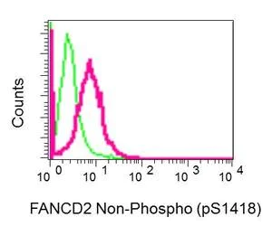
Flow Cytometry (Intracellular) - Anti-FANCD2 antibody [EPR2279(3)] (AB178705)
Intracellular flow cytometric analysis of permeabilized HeLa cells labeling FANCD2 with ab178705 at 1/10 dilution (red) compared toa rabbit IgG negative control (green).
- WB
Lab
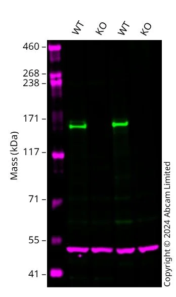
Western blot - Anti-FANCD2 antibody [EPR2279(3)] (AB178705)
Western blot : Anti-FANCD2 antibody [EPR2279(3)] (ab178705) staining at 1/1000 dilution, shown in green; Mouse anti-Alpha Tubulin [DM1A] (ab7291) loading control staining at 1/20000 dilution, shown in magenta. In Western blot, ab178705 was shown to bind specifically to FANCD2. A band was observed at 160 kDa in wild-type HCT 116 cell lysates with no signal observed at this size in FANCD2 knockout cell line. To generate this image, wild-type and FANCD2 knockout HCT 116 cell lysates were analysed. First, samples were run on an SDS-PAGE gel then transferred onto a nitrocellulose membrane. Membranes were blocked in 3 % milk in TBS-0.1 % Tween® 20 (TBS-T) before incubation with primary antibodies overnight at 4 °C. Blots were washed four times in TBS-T, incubated with secondary antibodies for 1 h at room temperature, washed again four times then imaged. Secondary antibodies used were Goat anti-Rabbit IgG H&L 800CW and Goat anti-Mouse IgG H&L 680RD at 1/20000 dilution.
All lanes:
Western blot - Anti-FANCD2 antibody [EPR2279(3)] (ab178705) at 1/1000 dilution
Lane 1:
Wild-type HCT 116 cell lysate at 20 µg
Lane 2:
FANCD2 knockout HCT 116 cell lysate at 20 µg
Lane 3:
Wild-type HeLa ab255448 cell lysate at 20 µg
Lane 4:
FANCD2 knockout HeLa <a href='/ko/products/cell-lines/human-fancd2-knockout-hela-cell-line-ab261743'>ab261743</a> cell lysate at 20 µg
Secondary
Lanes 1 - 4:
Goat anti-Rabbit IgG H&L 800CW at 1/20000 dilution
Lanes 1 - 4:
Goat anti-Mouse IgG H&L 680RD at 1/20000 dilution
Observed band size: 160 kDa
false
- WB
Lab
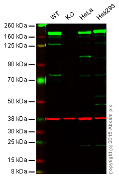
Western blot - Anti-FANCD2 antibody [EPR2279(3)] (AB178705)
Lanes 1 - 4 : Merged signal (red and green). Green - ab178705 observed at 166 kDa. Red - loading control, ab8245, observed at 37 kDa.
ab178705 was shown to specifically react with FANCD2 when FANCD2 knockout samples were used. Wild-type and FANCD2 knockout samples were subjected to SDS-PAGE. ab178705 and ab8245 (loading control to GAPDH) were diluted 1/1000 and 1/2000 respectively and incubated overnight at 4°C. Blots were developed with Goat anti-Rabbit IgG H&L (IRDye® 800CW) preadsorbed ab216773 and Goat anti-Mouse IgG H&L (IRDye® 680RD) preadsorbed ab216776 secondary antibodies at 1/10000 dilution for 1 h at room temperature before imaging.
All lanes:
Western blot - Anti-FANCD2 antibody [EPR2279(3)] (ab178705) at 1/1000 dilution
Lane 1:
Wild-type HAP1 cell lysate at 20 µg
Lane 2:
FANCD2 knockout HAP1 cell lysate at 20 µg
Lane 3:
HeLa cell lysate at 20 µg
Lane 4:
HEK293 cell lysate at 20 µg
Predicted band size: 166 kDa
false
- WB
Lab
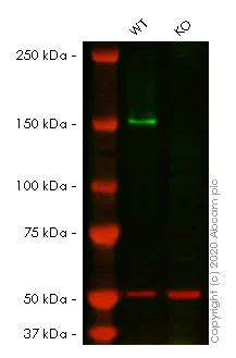
Western blot - Anti-FANCD2 antibody [EPR2279(3)] (AB178705)
Lanes 1- 2 : Merged signal (red and green). Green - ab178705 observed at 166 kDa. Red - Anti-alpha Tubulin antibody [DM1A] - Loading Control (ab7291) observed at 50 kDa.
ab178705 was shown to react with FANCD2 in wild-type HeLa cells in western blot. Loss of signal was observed when knockout cell line ab261743 (knockout cell lysate ab257173) was used. Wild-type HeLa and FANCD2 knockout HeLa cell lysates were subjected to SDS-PAGE. Membrane was blocked for 1 hour at room temperature in 0.1% TBST with 3% non-fat dried milk. ab178705 and Anti-alpha Tubulin antibody [DM1A] - Loading Control (ab7291) overnight at 4°C at a 1 in 1000 dilution and a 1 in 20000 dilution respectively. Blots were developed with Goat anti-Rabbit IgG H&L (IRDye®800CW) preadsorbed (ab216773) and Goat anti-Mouse IgG H&L (IRDye®680RD) preadsorbed (ab216776) secondary antibodies at 1 in 20000 dilution for 1 hour at room temperature before imaging.
All lanes:
Western blot - Anti-FANCD2 antibody [EPR2279(3)] (ab178705) at 1/1000 dilution
Lane 1:
Wild-type HeLa cell lysate at 20 µg
Lane 2:
FANCD2 knockout HeLa cell lysate at 20 µg
Lane 2:
Western blot - Human FANCD2 knockout HeLa cell line (<a href='/ko/products/cell-lines/human-fancd2-knockout-hela-cell-line-ab261743'>ab261743</a>)
Predicted band size: 166 kDa
Observed band size: 166 kDa
false
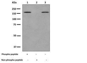
- WB
Supplier Data
Western blot - Anti-FANCD2 antibody [EPR2279(3)] (AB178705)
All lanes:
Western blot - Anti-FANCD2 antibody [EPR2279(3)] (ab178705) at 1/1000 dilution
Lane 1:
HeLa cell lysate at 10 µg with FANCD2 (pS1418) peptide
Lane 2:
HeLa cell lysate at 10 µg with Non-phospho peptide
Lane 3:
HeLa cell lysate at 10 µg
Predicted band size: 166 kDa
false
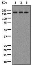
- WB
Supplier Data
Western blot - Anti-FANCD2 antibody [EPR2279(3)] (AB178705)
All lanes:
Western blot - Anti-FANCD2 antibody [EPR2279(3)] (ab178705) at 1/1000 dilution
Lane 1:
HeLa cell lysate untreated at 10 µg
Lane 2:
HeLa cell lysate treated with Alkaline Phosphatase at 10 µg
Lane 3:
HeLa cell lysate treated with Lambda Phosphatase at 10 µg
Predicted band size: 166 kDa
false
관련 conjugated 항체와 다양한 조성의 항체 (1)
-
Anti-FANCD2 antibody [EPR2279(3)] - BSA and Azide free
대체품
Primary Antibodies
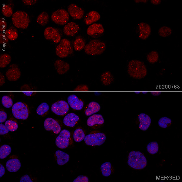
AB200763
Alexa Fluor® 647 Anti-FANCD2 antibody [EPR2302]
RabMAb|Recombinant|KO Validated
- 0
- 0
Primary Antibodies
AB109542
Anti-FANCD2 (phospho S1404) antibody [EPR2278(2)]
RabMAb|Recombinant
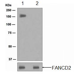
- 4
- 1
Primary Antibodies
AB108928
Anti-FANCD2 antibody [EPR2302]
20ul selling size|KO Validated|RabMAb|Recombinant
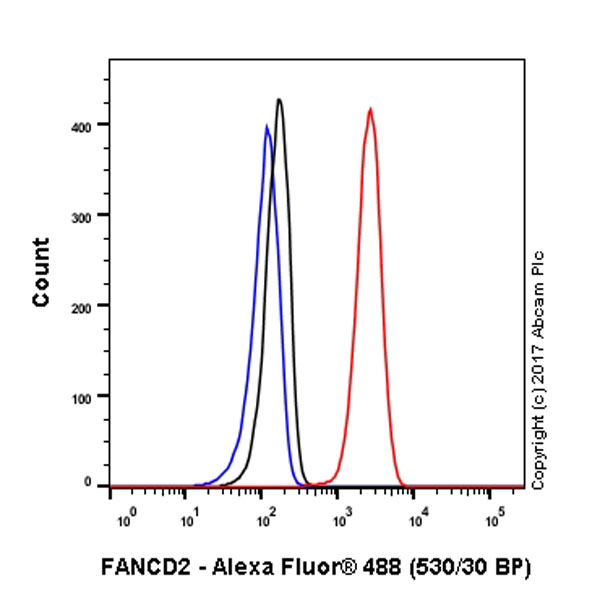
- 5
- 4
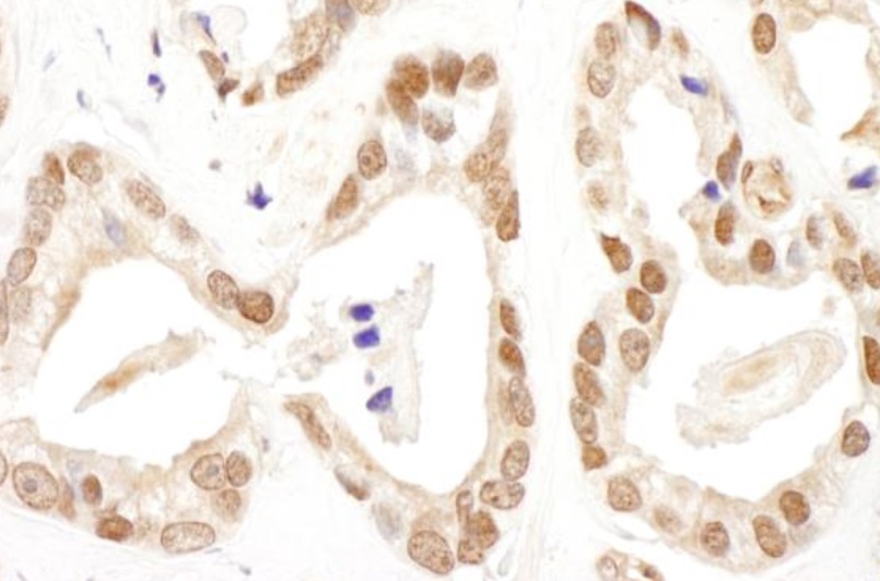
Primary Antibodies
AB200759
Alexa Fluor® 488 Anti-FANCD2 antibody [EPR2302]
RabMAb|Recombinant|KO Validated
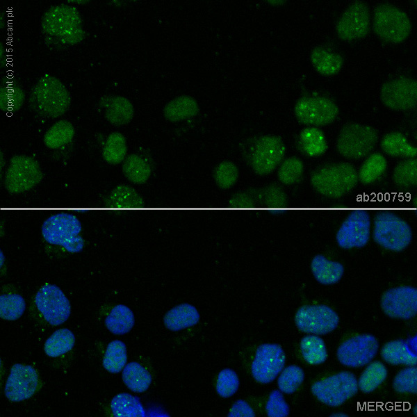
- 0
- 0
Primary Antibodies
AB221932
Anti-FANCD2 antibody [EPR2302] - BSA and Azide free
KO Validated|RabMAb|Recombinant
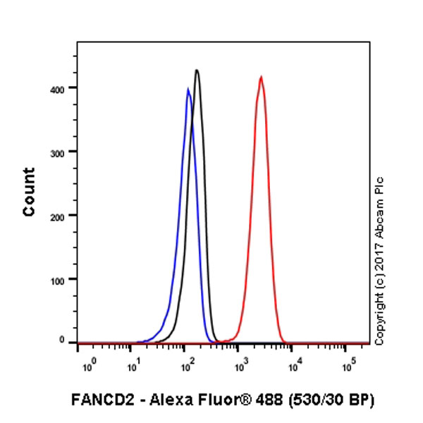
- 3
- 1
보완 제품

Primary Antibodies
AB133534
Anti-Rad51 antibody [EPR4030(3)]
20ul selling size|RabMAb|Recombinant
- 4
- 15
Primary Antibodies
AB181602
Anti-GAPDH antibody [EPR16891] - Loading Control
KD Validated|RabMAb|Recombinant|Lab Essentials|20ul selling size

- 5
- 23
Primary Antibodies
AB183734
RabMAb|Recombinant|Lab Essentials|20ul selling size
- 5
- 24
Reactivity 정보
제품 세부 정보
Species reactivity
Mouse, Rat: We have preliminary internal testing data to indicate this antibody may not react with these species.
Please contact us for more information.
Patented technology
Our RabMAb® technology is a patented hybridoma-based technology for making rabbit monoclonal antibodies. For details on our patents, please refer to RabMAb® patents.
What are the advantages of a recombinant monoclonal antibody?
This product is a recombinant monoclonal antibody, which offers several advantages including:
- - High batch-to-batch consistency and reproducibility
- - Improved sensitivity and specificity
- - Long-term security of supply
- - Animal-free batch production
For more information, read more on recombinant antibodies.
특성 및 보관 정보
제형
Purification 테크닉
보관 버퍼
배송 시 보관 조건
적절한 단기 보관 기간
적절한 단기 보관 조건
적절한 장기 보관 조건
분주 정보
보관 정보
추가 정보
This supplementary information is collated from multiple sources and compiled automatically.
Biological function summary
FANCD2 collaborates with multiple proteins as part of the Fanconi anemia (FA) complex to ensure genomic stability. The FA complex operates by facilitating the repair of DNA interstrand crosslinks that impede replication. FANCD2 becomes activated through monoubiquitination which serves as a signal for recruitment to DNA damage sites. It essentially functions as a coordinator that brings together important components for repair.
Pathways
FANCD2 integrates into the Fanconi anemia pathway and the homologous recombination repair pathway. These pathways are critical for maintaining DNA integrity and preventing chromosomal instability. Within these pathways interactions with proteins such as BRCA1 and BRCA2 reinforce the repair processes. FANCD2's connection to these proteins exemplifies its role in complex mechanisms that preserve genomic fidelity.
제품 프로토콜
- Visit the General protocols
- Visit the Troubleshooting
타겟 정보
대체 명칭 보기
제품이 사용된 논문 (4)
Recent publications for all applications. Explore the 전체 목록and refine your search
Cell death discovery 11:106 PubMed40091075
2025
Applications
Unspecified application
Species
Unspecified reactive species
Molecular cell 71:498-509.e4 PubMed30033371
2018
Applications
Unspecified application
Species
Unspecified reactive species
DNA repair 57:29-34 PubMed28648892
2017
Applications
Unspecified application
Species
Unspecified reactive species
Molecular cell 65:247-259 PubMed27986371
2016
Applications
Unspecified application
Species
Unspecified reactive species
Product promise
Please note: All products are 'FOR RESEARCH USE ONLY. NOT FOR USE IN DIAGNOSTIC OR THERAPEUTIC PROCEDURES'.
For licensing inquiries, please contact partnerships@abcam.com