FITC Anti-ATP5A antibody [15H4C4] - Mitochondrial Marker
- Lab Essentials
- 각 태그의 뜻
5
(1 리뷰)
|
(5 제품이 사용된 논문 )
- Flow Cyt (Intra)
Lab
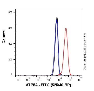
Flow Cytometry (Intracellular) - FITC Anti-ATP5A antibody [15H4C4] - Mitochondrial Marker (AB119688)
Flow cytometry overlay histogram showing HeLa cells stained with ab119688 (red line). The cells were fixed with 4% formaldehyde (10 min) and then permeabilised with 90% methanol for 30 min. The cells were incubated in 1x PBS containing 10% normal goat serum to block non-specific protein-protein interaction followed by the antibody (ab119688) (1x 10⁶ in 100μl at 1.0μg/ml) for 30min at 22°C.
Isotype control antibody (black line) was ab314952 (1.0μg/ml (1/500)) used at the same concentration and conditions as the primary antibody. Unlabelled sample (blue line) was also used as a control.
Acquisition of >5000 events were collected using a 50 mW Blue laser (488nm) and 525/40 bandpass filter.
- Flow Cyt (Intra)
Unknown
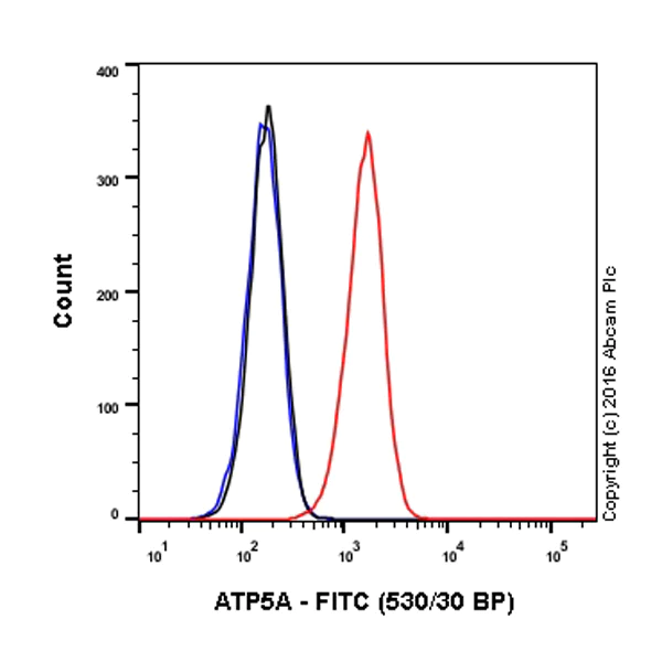
Flow Cytometry (Intracellular) - FITC Anti-ATP5A antibody [15H4C4] - Mitochondrial Marker (AB119688)
Overlay histogram showing HeLa cells stained with ab119688 (red line). The cells were fixed with 80% methanol (5 min) and then permeabilized with 0.1% PBS-Triton X-100 for 15 min. The cells were then incubated in 1x PBS / 10% normal goat serum to block non-specific protein-protein interactions followed by the antibody (ab119688, 1/100 dilution) for 30 min at 22°C.
Isotype control antibody (black line) was mouse IgG2b FITC (ab18419) used at the same concentration and conditions as the primary antibody. Unlabelled sample (blue line) was also used as a control.
Acquisition of >5,000 events were collected using a 50 mW Blue laser (488nm) and 530/30 bandpass filter.
This antibody gave a positive signal in HeLa cells fixed with 4% formaldehyde (10 min)/permeabilized with 0.1% PBS-Triton X-100 for 15 min used under the same conditions.
- ICC/IF
Unknown
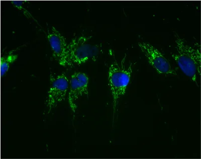
Immunocytochemistry/ Immunofluorescence - FITC Anti-ATP5A antibody [15H4C4] - Mitochondrial Marker (AB119688)
Mitochondrial localization of ATP5A using antibody ab119688. Cultured HeLa cells were fixed, permeabilized and then labeled with 15H4C4-FITC (1 µg/ml). Since the antibody is labeled with FITC no secondary antibody is necessary.
- ICC/IF
Lab
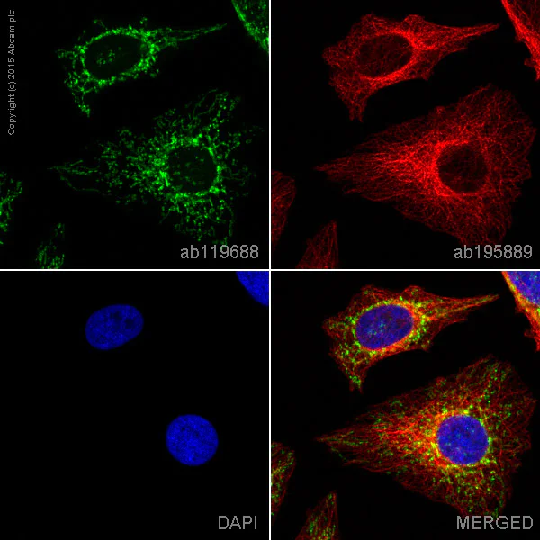
Immunocytochemistry/ Immunofluorescence - FITC Anti-ATP5A antibody [15H4C4] - Mitochondrial Marker (AB119688)
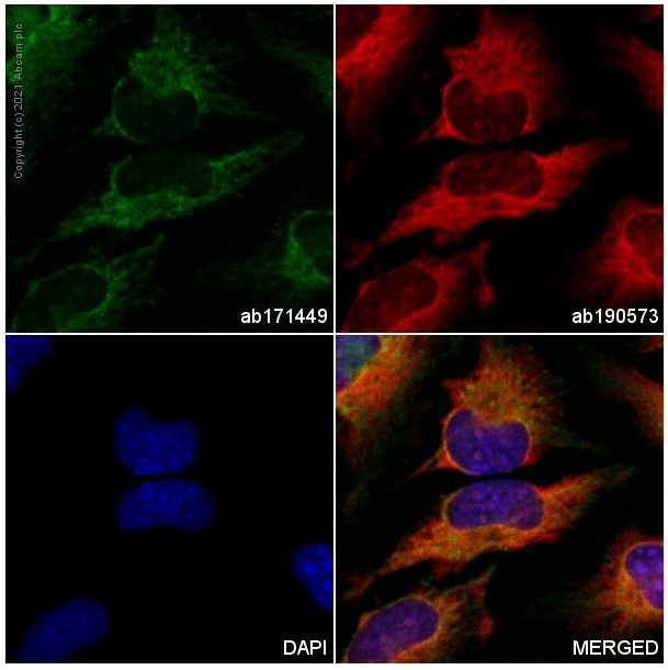
ab119688 staining ATP5A in HeLa cells. The cells were fixed with 4% formaldehyde (10 min), permeabilized with 0.1% Triton X-100 for 5 minutes and then blocked with 1% BSA/10% normal goat serum/0.3M glycine in 0.1% PBS-Tween for 1h. The cells were then incubated overnight at +4°C with ab119688 at 1/100 dilution (shown in green) and ab195889, Mouse monoclonal to alpha Tubulin (Alexa Fluor® 594), at 1/250 dilution (pseudocolored in red). Nuclear DNA was labelled with DAPI (shown in blue).
Image was taken with a confocal microscope (Leica-Microsystems, TCS SP8).
This product also gave a positive signal under the same testing conditions in HeLa cells fixed with 100% methanol (5min).
- Flow Cyt (Intra)
Unknown
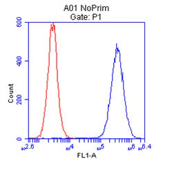
Flow Cytometry (Intracellular) - FITC Anti-ATP5A antibody [15H4C4] - Mitochondrial Marker (AB119688)
Overlay histogram showing HeLa cells stained with ab119688 (blue line). The cells were fixed with 4% paraformaldehyde (10 min) and then permeabilized with 0.1% PBS-Tween for 20 min. The cells were then incubated in 1x PBS / 10% normal goat serum / 0.3M glycine to block non-specific protein-protein interactions followed by the antibody ab119688 (15H4C4 FITC) at 1μg/1xE6/mL cells for 30 min at 22°C. Negative control (red line) is unstained d cells. Acquisition of >5,000 events was performed. This antibody gave a positive signal in HeLa cells fixed with 80% methanol (5 min)/permeabilized in 0.1% PBS-Tween used under the same conditions.
관련 conjugated 항체와 다양한 조성의 항체 (1)
-
Anti-ATP5A antibody [15H4C4] - Mitochondrial Marker
대체품
Primary Antibodies
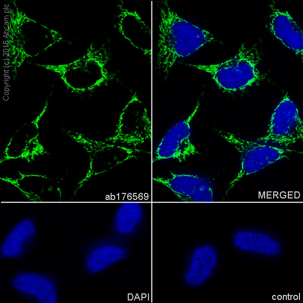
AB176569
Anti-ATP5A antibody [EPR13030(B)] - Mitochondrial Marker
RabMAb|Recombinant|Lab Essentials|20ul selling size
- 5
- 5
Primary Antibodies
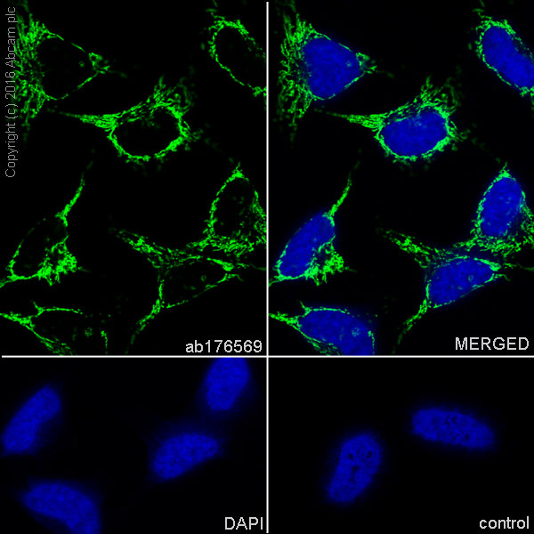
AB231692
Anti-ATP5A antibody [EPR13030(B)] - BSA and Azide free
RabMAb|Recombinant
- 5
- 2
Primary Antibodies
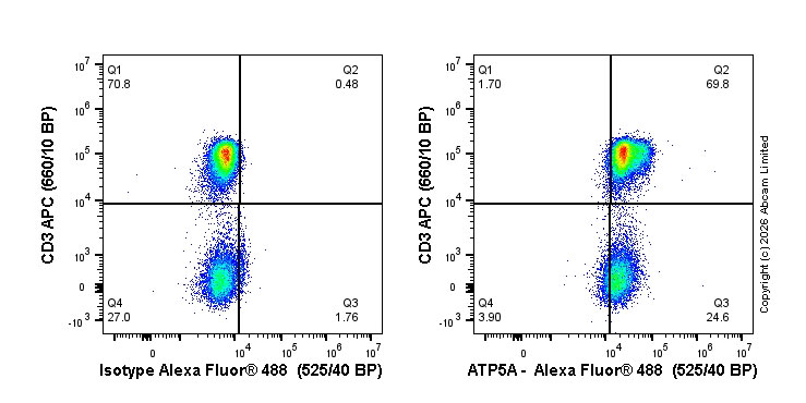
AB196467
Alexa Fluor® 488 Anti-ATP5A antibody [EPR13030(B)] - Mitochondrial Marker
RabMAb|Recombinant|Trial Size
- 0
- 0
Primary Antibodies
AB178421
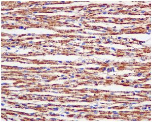
Anti-ATP5A antibody [EPR13031] - Mitochondrial Marker
Recombinant|Lab Essentials|RabMAb
- 0
- 0
Primary Antibodies
AB195891
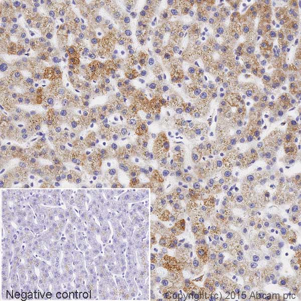
HRP Anti-ATP5A antibody [EPR13030(B)] - Mitochondrial Marker
BOND RX™ Validated|RabMAb|Recombinant
- 0
- 0
Primary Antibodies
AB216385
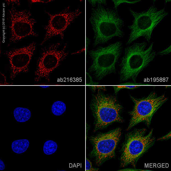
Alexa Fluor® 594 Anti-ATP5A antibody [EPR13030(B)] - Mitochondrial Marker
RabMAb|Recombinant
- 4
- 1
Primary Antibodies
AB196198
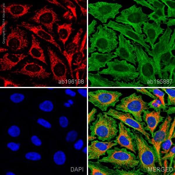
Alexa Fluor® 647 Anti-ATP5A antibody [EPR13030(B)] - Mitochondrial Marker
RabMAb|Recombinant|Trial Size
- 0
- 0
보완 제품
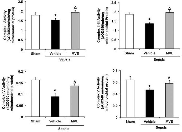
Primary Antibodies
AB212122
PE Anti-IDH2 antibody [EPR7577]
RabMAb|Recombinant|KO Validated|Trial Size
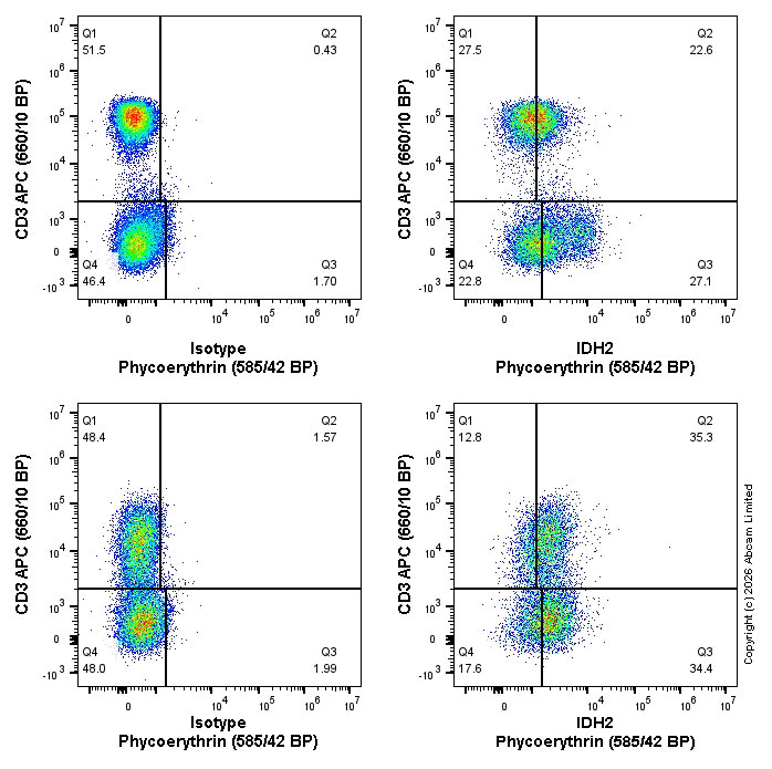
- 5
- 1
Primary Antibodies
AB197864
Alexa Fluor® 647 Anti-Hexokinase 1 antibody [EPR10134(B)] - Mitochondrial Outer Membrane Marker
RabMAb|Recombinant|KO Validated|Trial Size
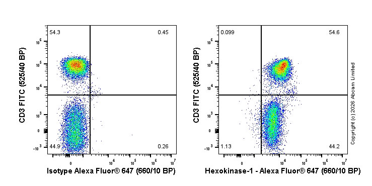
- 4
- 1
Primary Antibodies
AB210451
PE Anti-ASS1 antibody [EPR12398]
RabMAb|Recombinant|KO Validated|Trial Size
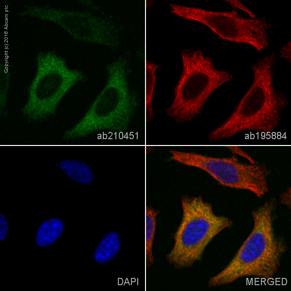
- 4
- 1
Reactivity 정보
특성 및 보관 정보
제형
Purification 테크닉
Purification 관련 사항
보관 버퍼
배송 시 보관 조건
적절한 단기 보관 기간
적절한 단기 보관 조건
적절한 장기 보관 조건
보관 정보
추가 정보
This supplementary information is collated from multiple sources and compiled automatically.
Biological function summary
ATP5A is essential in cellular respiration serving as a catalytic core of the F1 component of ATP synthase. As part of the multi-subunit enzyme complex ATP synthase is responsible for ATP production the primary energy currency in cells. The ATP5A subunit works in conjunction with other subunits of the enzyme oligomer to facilitate the conversion of energy released during oxidative phosphorylation into a usable form. The protein's efficiency in this biological role underpins its importance in sustaining cellular energy homeostasis.
Pathways
ATP5A plays a pivotal role in oxidative phosphorylation and the electron transport chain integral components of cellular respiration. The oxidative phosphorylation pathway depends on this protein to manage the synthesis of ATP molecules while the electron transport chain creates the proton gradient necessary for ATP production. ATP5A is functionally connected to other proteins in these pathways such as ATP5B and cytochrome c oxidase working in a coordinated manner to ensure efficient energy transfer and maintenance.
제품 프로토콜
- Visit the General protocols
- Visit the Troubleshooting
타겟 정보
대체 명칭 보기
제품이 사용된 논문 (5)
Recent publications for all applications. Explore the 전체 목록and refine your search
Journal of cell science 135: PubMed35833493
2022
Applications
Unspecified application
Species
Unspecified reactive species
Nature 591:471-476 PubMed33627869
2021
Applications
Unspecified application
Species
Unspecified reactive species
Frontiers in cell and developmental biology 7:305 PubMed31929983
2019
Applications
Unspecified application
Species
Unspecified reactive species
Current biology : CB 27:1928-1940.e6 PubMed28669756
2017
Applications
Unspecified application
Species
Unspecified reactive species
Nature methods 11:861-7 PubMed24930129
2014
Applications
Unspecified application
Species
Unspecified reactive species
Product promise
Please note: All products are 'FOR RESEARCH USE ONLY. NOT FOR USE IN DIAGNOSTIC OR THERAPEUTIC PROCEDURES'.
For licensing inquiries, please contact partnerships@abcam.com