Anti-Glycam1 antibody [EPR30103-507]
- Advanced Validation
- RabMAb
- Recombinant
- 20ul selling size
- 각 태그의 뜻
0
(1 리뷰)
|
(0 출판물)
- IHC-Fr
Lab
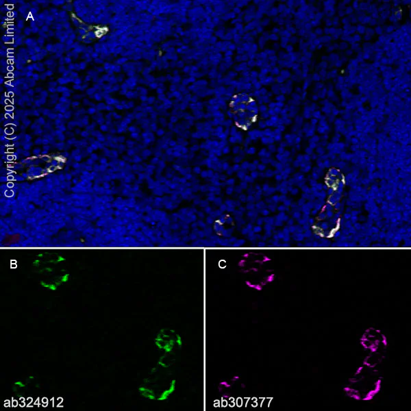
Immunohistochemistry (Frozen sections) - Anti-Glycam1 antibody [EPR30103-507] (AB324912)
Immunohistochemical analysis of 4% PFA-fixed, 0.2% Triton X-100 permeabilized frozen Mouse lymph node (fresh frozen) tissue labeling Glycam1 with ab324912 at 1/500 (1.01 μg/ml) dilution followed by ab150081 Goat Anti-Rabbit IgG H&L (Alexa Fluor® 488) preadsorbed at 1/1000 (2 ug/mL) dilution (Green). ab307377 was used as a co-localization positive control staining high endothelial venules (magenta).
Panel A : merged staining of anti-GLYCAM1 (ab324912, green), anti-VWF (ab307377, magenta) on mouse lymph node.
Panel B : anti-GLYCAM1 stained on mouse lymph node.
Panel C : anti-VWF stained in high endothelial venules (HEV) of mouse lymph node.
The section was incubated in two rounds of staining : in the order of ab324912 and ab307377, for 1 hr at room temperature. The nuclear counterstain was DAPI (Blue). The section was then mounted using Fluoromount®.
Heat mediated antigen retrieval using sodium citrate buffer (10mM citrate pH 6.0 + 0.05% Tween-20)
The immunostaining was performed on a Leica Biosystems BOND® RX instrument. Image was taken with a confocal microscope (Leica-Microsystems, TCS SP8).
- IHC-Fr
Lab
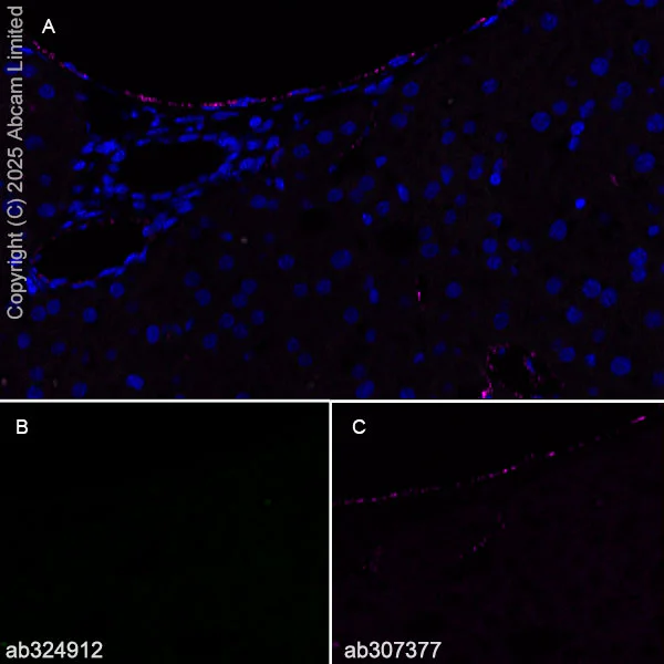
Immunohistochemistry (Frozen sections) - Anti-Glycam1 antibody [EPR30103-507] (AB324912)
Immunohistochemical analysis of 4% PFA-fixed, 0.2% Triton X-100 permeabilized frozen Mouse liver (perfused fixed-fresh frozen) tissue, negative control, labeling Glycam1 with ab324912 at 1/500 (1.01 μg/ml) dilution followed by ab150081 Goat Anti-Rabbit IgG H&L (Alexa Fluor® 488) preadsorbed at 1/1000 (2 ug/mL) dilution (Green). ab307377 was used as a co-localization positive control (magenta).
Panel A : merged staining of anti-GLYCAM1 (ab324912, green), anti-VWF (ab307377, magenta) on mouse liver.
Panel B : anti-GLYCAM1 stained on mouse liver.
Panel C : anti-VWF stained in endothelial cells in mouse liver.
The section was incubated in two rounds of staining : in the order of ab324912 and ab307377, for 1 hr at room temperature. The nuclear counterstain was DAPI (Blue). The section was then mounted using Fluoromount®.
Heat mediated antigen retrieval using sodium citrate buffer (10mM citrate pH 6.0 + 0.05% Tween-20)
The immunostaining was performed on a Leica Biosystems BOND® RX instrument. Image was taken with a confocal microscope (Leica-Microsystems, TCS SP8).
- mIHC
Lab
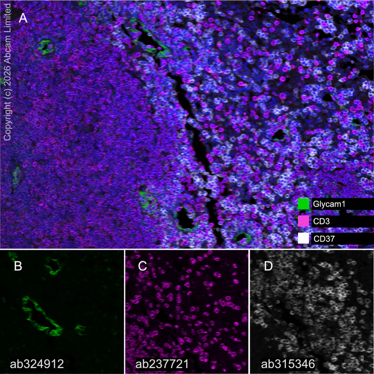
Multiplex immunohistochemistry - Anti-Glycam1 antibody [EPR30103-507] (AB324912)
Multiplex immunohistochemistry analysis of formalin/PFA-fixed paraffin-embedded mouse lymph node tissue staining Glycam1 with ab324912 at a 1/4000 ( 0.126 μg/ml) dilution, ab237721 anti-CD3 used at 1/2000 dilution ( 0.26 μg/ml) and ab315346 anti-CD37 used at 1/2000 dilution (0.257 μg/ml).
Panel A : anti-Glycam1 (green; Opal™520), anti-CD3 (magenta; Opal™690), anti-CD37 (gray; Opal™570) on mouse lymph node.
Panel B : anti-Glycam1 staining HEV (highly endothelial venules) in mouse lymph node.
Panel C : anti-CD3 staining T lymphocytes in mouse lymph node.
Panel D : anti-CD37 staining B lymphocytes in mouse lymph node.
Nuclear DNA was labeled with DAPI (shown in blue).
The section was incubated in three rounds of staining : in the order of ab324912, ab237721 and ab315346 for 30 mins at room temperature. Each round was followed by a separate fluorescent tyramide signal amplification system.
The immunostaining was performed on a Leica Biosystems BOND® RX instrument with an Opal™ 4-color kit. Image acquisition was performed with Leica SP8 confocal microscope.
Heat mediated antigen retrieval was performed with Tris-EDTA buffer (pH 9.0, Epitope Retrieval Solution2) for 20 mins.
- mIHC
Lab
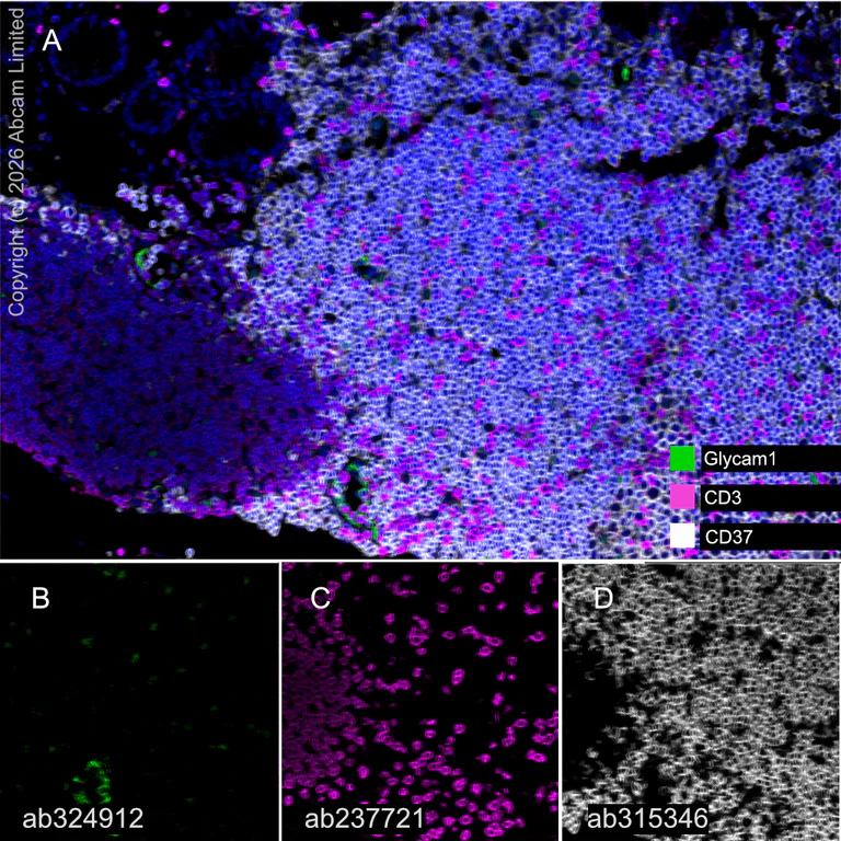
Multiplex immunohistochemistry - Anti-Glycam1 antibody [EPR30103-507] (AB324912)
Multiplex immunohistochemistry analysis of formalin/PFA-fixed paraffin-embedded mouse colon tissue staining Glycam1 with ab324912 at a 1/4000 ( 0.126 μg/ml) dilution, ab237721 anti-CD3 used at 1/2000 dilution ( 0.26 μg/ml) and ab315346 anti-CD37 used at 1/2000 dilution (0.257 μg/ml).
Panel A : anti-Glycam1 (green; Opal™520), anti-CD3 (magenta; Opal™690), anti-CD37 (gray; Opal™570) on mouse colon.
Panel B : anti-Glycam1 staining HEV (highly endothelial venules) in mouse colon.
Panel C : anti-CD3 staining T lymphocytes in mouse colon.
Panel D : anti-CD37 staining B lymphocytes in mouse colon.
Nuclear DNA was labeled with DAPI (shown in blue).
The section was incubated in three rounds of staining : in the order of ab324912, ab237721 and ab315346 for 30 mins at room temperature. Each round was followed by a separate fluorescent tyramide signal amplification system.
The immunostaining was performed on a Leica Biosystems BOND® RX instrument with an Opal™ 4-color kit. Image acquisition was performed with Leica SP8 confocal microscope.
Heat mediated antigen retrieval was performed with Tris-EDTA buffer (pH 9.0, Epitope Retrieval Solution2) for 20 mins.
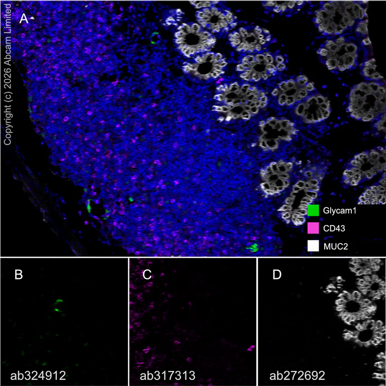
- mIHC
Lab
Multiplex immunohistochemistry - Anti-Glycam1 antibody [EPR30103-507] (AB324912)
Multiplex immunohistochemistry analysis of formalin/PFA-fixed paraffin-embedded mouse colon tissue staining Glycam1 with ab324912 at a 1/4000 ( 0.126 μg/ml) dilution, ab317313 anti-CD43 used at 1/500 dilution (1.052 μg/ml) and ab272692 anti-MUC2 used at 1/2000 dilution (0.26 μg/ml).
Panel A : anti-Glycam1 (green; Opal™520), anti-CD43 (magenta; Opal™690), anti-MUC2 (gray; Opal™570) on mouse colon.
Panel B : anti-Glycam1 staining HEV (highly endothelial venules) in mouse colon.
Panel C : anti-CD43 staining T lymphocytes in mouse colon.
Panel D : anti-MUC2 staining goblet cells in mouse colon.
Nuclear DNA was labeled with DAPI (shown in blue).
The section was incubated in three rounds of staining : in the order of ab324912, ab237721 and ab315346 for 30 mins at room temperature. Each round was followed by a separate fluorescent tyramide signal amplification system.
The immunostaining was performed on a Leica Biosystems BOND® RX instrument with an Opal™ 4-color kit. Image acquisition was performed with Leica SP8 confocal microscope.
Heat mediated antigen retrieval was performed with Tris-EDTA buffer (pH 9.0, Epitope Retrieval Solution2) for 20 mins.
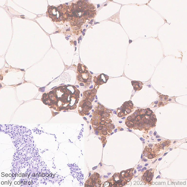
- IHC-P
Supplier Data
Immunohistochemistry (Formalin/PFA-fixed paraffin-embedded sections) - Anti-Glycam1 antibody [EPR30103-507] (AB324912)
Immunohistochemical analysis of paraffin-embedded Mouse breast tissue labeling Glycam1 with ab324912 at 1/4000 (0.126 ug/ml) dilution, followed by a ready to use LeicaDS9800 (Bond™ Polymer Refine Detection).
Positive staining on mouse breast. The primary antibody was incubated for 30 mins at room temperature.
The immunostaining was performed on a Leica Biosystems BOND® RX instrument. Incubate slides with 3% Hydrogen Peroxide for 10 mins at room temperature after secondary antibody incubation to reduce the background.
Counterstained with Hematoxylin.
Secondary antibody only control : Secondary antibody is a ready to use LeicaDS9800 (Bond™ Polymer Refine Detection).
Heat mediated antigen retrieval was performed with Tris-EDTA buffer (pH 9.0, Epitope Retrieval Solution2) for 20 mins.
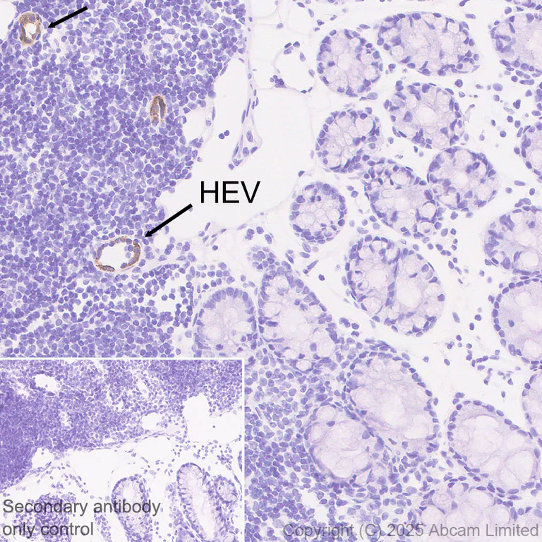
- IHC-P
Supplier Data
Immunohistochemistry (Formalin/PFA-fixed paraffin-embedded sections) - Anti-Glycam1 antibody [EPR30103-507] (AB324912)
Immunohistochemical analysis of paraffin-embedded Mouse colon tissue labeling Glycam1 with ab324912 at 1/4000 (0.126 ug/ml) dilution, followed by a ready to use LeicaDS9800 (Bond™ Polymer Refine Detection).
Positive staining on HEV (highly endothelial venules) in mouse colon. The primary antibody was incubated for 30 mins at room temperature.
The immunostaining was performed on a Leica Biosystems BOND® RX instrument. Incubate slides with 3% Hydrogen Peroxide for 10 mins at room temperature after secondary antibody incubation to reduce the background.
Counterstained with Hematoxylin.
Secondary antibody only control : Secondary antibody is a ready to use LeicaDS9800 (Bond™ Polymer Refine Detection).
Heat mediated antigen retrieval was performed with Tris-EDTA buffer (pH 9.0, Epitope Retrieval Solution2) for 20 mins
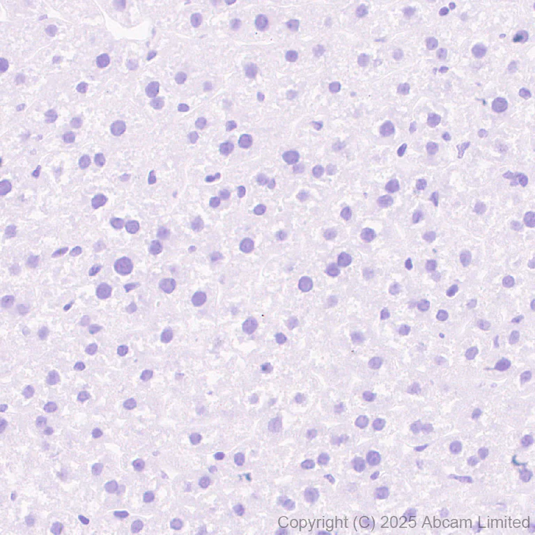
- IHC-P
Supplier Data
Immunohistochemistry (Formalin/PFA-fixed paraffin-embedded sections) - Anti-Glycam1 antibody [EPR30103-507] (AB324912)
Immunohistochemical analysis of paraffin-embedded Mouse liver tissue labeling Glycam1 with ab324912 at 1/4000 (0.126 ug/ml) dilution, followed by a ready to use LeicaDS9800 (Bond™ Polymer Refine Detection).
Negative control : no staining on mouse liver. The primary antibody was incubated for 30 mins at room temperature.
The immunostaining was performed on a Leica Biosystems BOND® RX instrument. Incubate slides with 3% Hydrogen Peroxide for 10 mins at room temperature after secondary antibody incubation to reduce the background.
Counterstained with Hematoxylin.
Secondary antibody only control : Secondary antibody is a ready to use LeicaDS9800 (Bond™ Polymer Refine Detection).
Heat mediated antigen retrieval was performed with Tris-EDTA buffer (pH 9.0, Epitope Retrieval Solution2) for 20 mins
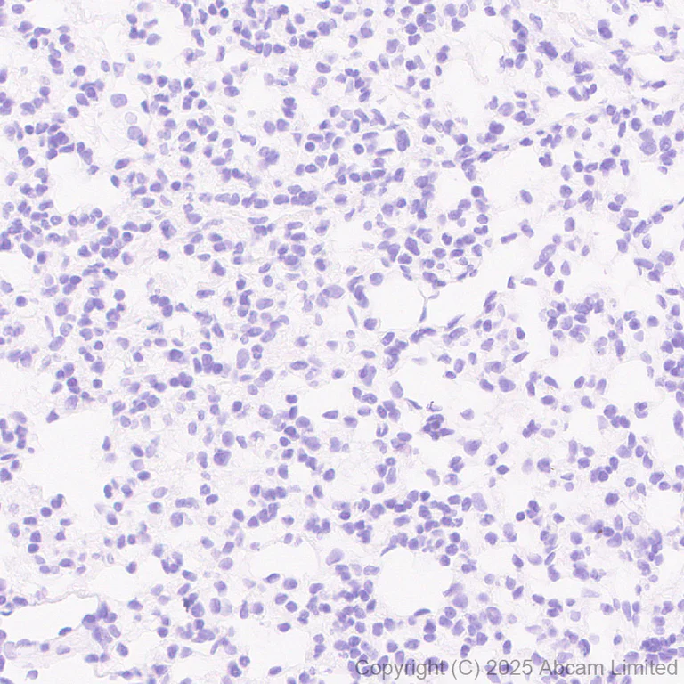
- IHC-P
Supplier Data
Immunohistochemistry (Formalin/PFA-fixed paraffin-embedded sections) - Anti-Glycam1 antibody [EPR30103-507] (AB324912)
Immunohistochemical analysis of paraffin-embedded Mouse lung tissue labeling Glycam1 with ab324912 at 1/4000 (0.126 ug/ml) dilution, followed by a ready to use LeicaDS9800 (Bond™ Polymer Refine Detection).
Negative control : no staining on mouse lung. The primary antibody was incubated for 30 mins at room temperature.
The immunostaining was performed on a Leica Biosystems BOND® RX instrument. Incubate slides with 3% Hydrogen Peroxide for 10 mins at room temperature after secondary antibody incubation to reduce the background.
Counterstained with Hematoxylin.
Secondary antibody only control : Secondary antibody is a ready to use LeicaDS9800 (Bond™ Polymer Refine Detection).
Heat mediated antigen retrieval was performed with Tris-EDTA buffer (pH 9.0, Epitope Retrieval Solution2) for 20 mins
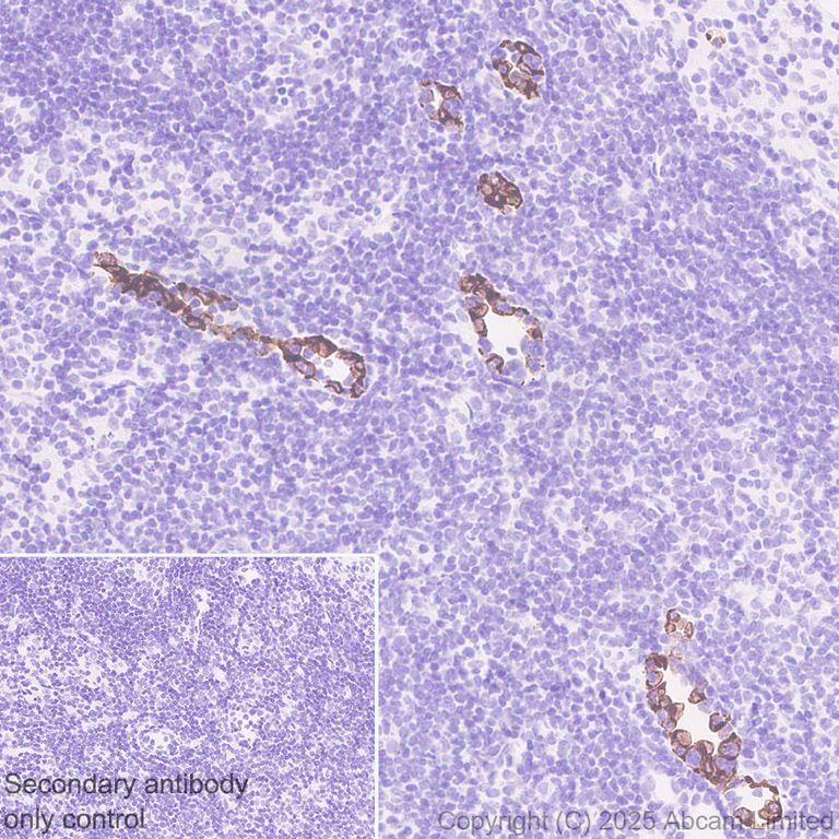
- IHC-P
Supplier Data
Immunohistochemistry (Formalin/PFA-fixed paraffin-embedded sections) - Anti-Glycam1 antibody [EPR30103-507] (AB324912)
Immunohistochemical analysis of paraffin-embedded Mouse lymph node tissue labeling Glycam1 with ab324912 at 1/4000 (0.126 ug/ml) dilution, followed by a ready to use LeicaDS9800 (Bond™ Polymer Refine Detection).
Positive staining on HEV (highly endothelial venules) of mouse lymph node. The primary antibody was incubated for 30 mins at room temperature.
The immunostaining was performed on a Leica Biosystems BOND® RX instrument. Incubate slides with 3% Hydrogen Peroxide for 10 mins at room temperature after secondary antibody incubation to reduce the background.
Counterstained with Hematoxylin.
Secondary antibody only control : Secondary antibody is a ready to use LeicaDS9800 (Bond™ Polymer Refine Detection).
Heat mediated antigen retrieval was performed with Tris-EDTA buffer (pH 9.0, Epitope Retrieval Solution2) for 20 mins
보완 제품
Primary Antibodies
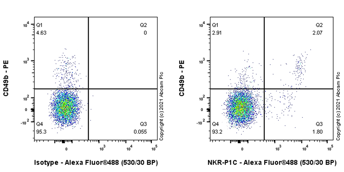
AB289542
Anti-NKR-P1C/NK1.1 antibody [EPR22990-31]
20ul selling size|RabMAb|Recombinant
- 4
- 2


Primary Antibodies
AB282575
Anti-ICAM1 antibody [EPR24639-3]
BOND RX™ Validated|20ul selling size|RabMAb|Recombinant|KO Validated

- 5
- 1
Reactivity 정보
제품 세부 정보
Patented technology
Our RabMAb® technology is a patented hybridoma-based technology for making rabbit monoclonal antibodies. For details on our patents, please refer to RabMAb® patents.
What are the advantages of a recombinant monoclonal antibody?
This product is a recombinant monoclonal antibody, which offers several advantages including:
- - High batch-to-batch consistency and reproducibility
- - Improved sensitivity and specificity
- - Long-term security of supply
- - Animal-free batch production
For more information, read more on recombinant antibodies.
특성 및 보관 정보
제형
Purification 테크닉
보관 버퍼
배송 시 보관 조건
적절한 단기 보관 기간
적절한 단기 보관 조건
적절한 장기 보관 조건
분주 정보
보관 정보
제품 프로토콜
- Visit the General protocols
- Visit the Troubleshooting
타겟 정보
대체 명칭 보기
Product promise
Please note: All products are 'FOR RESEARCH USE ONLY. NOT FOR USE IN DIAGNOSTIC OR THERAPEUTIC PROCEDURES'.
For licensing inquiries, please contact partnerships@abcam.com