Anti-HDAC2 antibody [HDAC2-62]
5
(6 리뷰들)
|
(87 제품이 사용된 논문 )
Mouse Monoclonal HDAC2 antibody. Suitable for IP, WB and reacts with Human, Mouse samples. Cited in 87 publications. Immunogen corresponding to Synthetic Peptide within Human HDAC2 aa 450 to C-terminus conjugated to Keyhole Limpet Haemocyanin.
대체 명칭 보기
Histone deacetylase 2, HD2, Protein deacylase HDAC2, HDAC2

- IP
Supplier Data
Immunoprecipitation - Anti-HDAC2 antibody [HDAC2-62] (AB12169)
Western Blot using Rabbit anti-HDAC2
Lane 1:
Immunoprecipitation - Anti-HDAC2 antibody [HDAC2-62] (ab12169) at 5 µg
Lane 2:
Immunoprecipitation - Anti-HDAC2 antibody [HDAC2-62] (ab12169) at 2.5 µg
Lane 3:
Negative Control at 5 µg
All lanes:
HeLa whole cell extract
Predicted band size: 55 kDa
false

- WB
Supplier Data
Western blot - Anti-HDAC2 antibody [HDAC2-62] (AB12169)
All lanes:
Western blot - Anti-HDAC2 antibody [HDAC2-62] (ab12169) at 0.5 µg/mL
Lane 1:
Hek293T cell Lysate
Lane 2:
HeLa cell Lysate
Lane 3:
Jurkat cell Lysate
Lane 4:
K562 cell Lysate
Lane 5:
Neuro-2a cell Lysate
Lane 6:
NIH-3T3 cell Lysate
Secondary
All lanes:
Goat Anti-Mouse IgG-Peroxidase
Predicted band size: 55 kDa
false

- WB
AbReview24155****
Western blot - Anti-HDAC2 antibody [HDAC2-62] (AB12169)
All lanes:
Western blot - Anti-HDAC2 antibody [HDAC2-62] (ab12169) at 1/250 dilution
All lanes:
Human glioblastoma cell line at 120 µg
Secondary
All lanes:
HRP-conjugated Goat anti-Mouse at 1/10000 dilution
Predicted band size: 55 kDa
Observed band size: 45 kDa,55 kDa
true
Exposure time: 5min
This image is courtesy of an anonymous abreview.
- WB
CiteAb
Western blot - Anti-HDAC2 antibody [HDAC2-62] (AB12169)
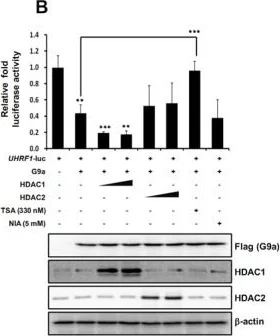
Western Blotting using Anti-HDAC2 antibody [HDAC2-62], ab12169. Publication image from Kook, H. et al., 2015, Nucleic Acids Res, 25765655. Legend direct from paper.
G9a-mediated transcriptional repression of UHRF1 is YY1 dependent. (A) 293T cells were cotransfected with the pGL3-UHRF1 promoter, Flag-G9a and Flag-YY1. Following transfection, cells were grown for 48 h, and cell extracts were assayed for luciferase activity. Expression of the transfected constructs is shown in the immunoblot analysis. (B) pGL3-UHRF1 promoter and the indicated constructs were cotransfected into 293T cells. Twenty-four hours after transfection, 330 nM TSA or 5 mM NIA was added for 24 h, and luciferase activities were subsequently measured. G9a, HDAC1 and HDAC2 expression was confirmed by immunoblot analysis. (C) 293T cells were cotransfected with the pGL3-UHRF1 promoter, Flag-G9a, siCTL RNA and siYY1 RNAs (100 nM). Luciferase activity was measured 48 h after transfection. G9a overexpression and YY1 knockdown by two different siYY1 RNAs are shown in the immunoblot analysis. (A–C) Luciferase activity was normalized to that of β-galactosidase, and the results are presented as means ± SD (n = 3). *P < 0.05, **P < 0.01, ***P < 0.001. (D) 293T cells were transfected with siCTL RNA or siYY1 RNAs (100 nM). YY1, G9a, and UHRF1 expression levels were confirmed using real time-PCR and immunoblot analysis. YY1 knockdown by siYY1 RNA is shown in the immunoblot analysis. All results are representative of at least three independent experiments (±SDs). **P < 0.01, ***P < 0.001. (E) 293T cells were transfected with siCTL RNA or siYY1 RNAs. ChIP analysis was performed using anti-G9a, anti-YY1, and anti-H3K9-me2 antibodies, and the results were confirmed by real-time PCR. Recruitment of G9a, YY1 and H3K9-me2 to the UHRF1 promoter and distal region was normalized by input. All results represent at least three independent experiments (±SDs). *P < 0.05, **P < 0.01, ***P < 0.001.
false
- WB
CiteAb
Western blot - Anti-HDAC2 antibody [HDAC2-62] (AB12169)
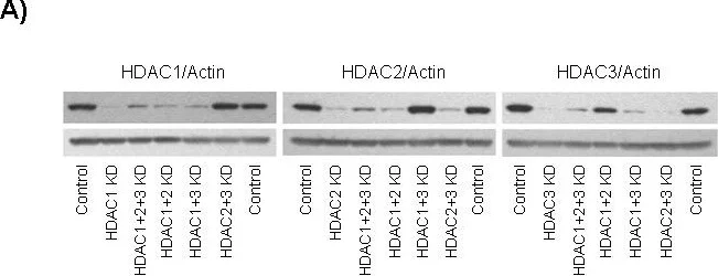
Western Blotting using Anti-HDAC2 antibody [HDAC2-62], ab12169. Publication image from Sehested, M. et al., 2008, Mol Cancer, 18789133. Legend direct from paper.
Effects of HDAC depletion by siRNA in HeLa cells. A) Down-regulation of HDAC1, 2 or 3 protein after treatment with HDAC1, 2 or 3-targeted siRNA respectively, alone or in combination with other siRNAs at 72 hours post-transfection. Top-row is the appropriate antibody (HDAC1, 2 or 3), while the bottom-row is loading control actin; B) viability as measured by ATP present in culture. For each HDAC knockdown condition, viability was consistently reduced as compared to scrambled siRNA control, in independent measurements (p < 0.01, n = 24); C) The activity of effector caspases 3 and 7 corresponding to apoptotic progression upon HDAC down-regulation or belinostat treatment at increasing concentrations for 24 hours for comparison; D) flow cytometric analysis of DNA content as a function of cell number in control or HDAC combination KD at 48 hours post-transfection, or belinostat treatment at 5.0 µM for 24 hours.
false
대체품

Primary Antibodies
AB196518
Alexa Fluor® 647 Anti-HDAC2 antibody [Y461]
RabMAb|Recombinant|KO Validated

- 0
- 0
Primary Antibodies
AB124974
Anti-HDAC2 antibody [EPR5001] - ChIP Grade
20ul selling size|RabMAb|Recombinant|KO Validated

- 4
- 4
Primary Antibodies
AB32117
20ul selling size|BOND RX™ Validated|RabMAb|Advanced Validation|Recombinant|KO Validated

- 5
- 12
Primary Antibodies
AB213700
Anti-HDAC2 antibody [Y461] - BSA and Azide free
BOND RX™ Validated|RabMAb|Advanced Validation|Recombinant|KO Validated

- 0
- 0
Primary Antibodies
AB219053
Anti-HDAC2 antibody [EPR20117]
Recombinant|KO Validated|Advanced Validation|RabMAb|20ul selling size

- 0
- 0
Primary Antibodies
AB196471
Alexa Fluor® 488 Anti-HDAC2 antibody [Y461]
RabMAb|Recombinant|KO Validated

- 0
- 0
보완 제품

Primary Antibodies
AB1791
Anti-Histone H3 antibody - Nuclear Marker and ChIP Grade
Lab Essentials

- 5
- 233


Primary Antibodies
AB183734
RabMAb|Recombinant|Lab Essentials|20ul selling size

- 5
- 24
Reactivity 정보
특성 및 보관 정보
제형
Purification 테크닉
보관 버퍼
배송 시 보관 조건
적절한 단기 보관 기간
적절한 단기 보관 조건
적절한 장기 보관 조건
분주 정보
보관 정보
추가 정보
This supplementary information is collated from multiple sources and compiled automatically.
Biological function summary
HDAC2 is involved in cell cycle regulation and differentiation. It is part of the co-repressor complex and interacts with other proteins such as Sin3 and NuRD to repress transcription. These interactions are vital for maintaining normal cellular functions and ensuring proper response to external signals. This protein is also connected to HEK293T cells which are a common model in scientific studies due to their robust expression of proteins like HDAC2.
Pathways
HDAC2 participates in the regulation of transcriptional activity and cellular stress response. It is an important player in the histone modification pathway interacting with proteins such as HDAC1 and transcriptional regulators like REST. HDAC2 is also linked with the Notch signaling pathway which is essential for cell fate decisions. These pathways highlight its role in both maintaining cellular homeostasis and adapting to changes in the cellular environment.
제품 프로토콜
- Visit the General protocols
- Visit the Troubleshooting
타겟 정보
제품이 사용된 논문 (87)
Recent publications for all applications. Explore the 전체 목록 and refine your search
Nature biotechnology 43:63-75 PubMed38459338
2024
Applications
Unspecified application
Species
Unspecified reactive species
Cell reports 43:113778 PubMed38341854
2024
Applications
Unspecified application
Species
Unspecified reactive species
Acta biochimica et biophysica Sinica 56:62-70 PubMed38143381
2023
Applications
Unspecified application
Species
Unspecified reactive species
Frontiers in molecular neuroscience 16:1207911 PubMed37389091
2023
Applications
Unspecified application
Species
Unspecified reactive species
Nature communications 14:1521 PubMed36934083
2023
Applications
Unspecified application
Species
Unspecified reactive species
SLAS discovery : advancing life sciences R & D 27:440-447 PubMed36240996
2022
Applications
Unspecified application
Species
Unspecified reactive species
American journal of translational research 14:6137-6149 PubMed36247238
2022
Applications
Unspecified application
Species
Unspecified reactive species
The Journal of neuroscience : the official journal of the Society for Neuroscience 42:4594-4604 PubMed35501151
2022
Applications
Unspecified application
Species
Unspecified reactive species
Cell reports 38:110534 PubMed35320709
2022
Applications
Unspecified application
Species
Unspecified reactive species
Journal of experimental & clinical cancer research : CR 41:34 PubMed35073946
2022
Applications
Unspecified application
Species
Unspecified reactive species
Product promise
Please note: All products are 'FOR RESEARCH USE ONLY. NOT FOR USE IN DIAGNOSTIC OR THERAPEUTIC PROCEDURES'.
For licensing inquiries, please contact partnerships@abcam.com