Anti-Iba1 antibody [EPR16588] - Goat IgG (Chimeric) - BSA and Azide free
- BOND RX™ Validated
- Recombinant
- 각 태그의 뜻
Be the first to review this product! 리뷰 제출
|
(0 출판물)

- IHC-P
Lab
Immunohistochemistry (Formalin/PFA-fixed paraffin-embedded sections) - Anti-Iba1 antibody [EPR16588] - Goat IgG (Chimeric) - BSA and Azide free (AB289876)
This data was developed using ab289874, the same antibody clone in a different buffer formulation.
Immunohistochemical analysis of paraffin-embedded Human liver tissue labeling Iba1 with ab289874 at 1/1000 dilution, followed by a ready to use LeicaDS9800 (Bond™ Polymer Refine Detection).
Positive staining on Kupffer cells in the human liver. The section was incubated with ab289874 for 30 mins at room temperature and followed by specific anti-goat IgG antibody (ab97101) for 8 mins. The immunostaining was performed on a Leica Biosystems BOND®RX instrument.
Counterstained with Hematoxylin.
Secondary antibody only control : Secondary antibody is a ready to use LeicaDS9800 (Bond™ Polymer Refine Detection).
Heat mediated antigen retrieval with Tris-EDTA buffer (pH 9.0, epitope retrieval solution2) for 20 mins.

- IHC-P
Lab
Immunohistochemistry (Formalin/PFA-fixed paraffin-embedded sections) - Anti-Iba1 antibody [EPR16588] - Goat IgG (Chimeric) - BSA and Azide free (AB289876)
This data was developed using ab289874, the same antibody clone in a different buffer formulation.
Immunohistochemical analysis of paraffin-embedded Human cerebrum tissue labeling Iba1 with ab289874 at 1/1000 dilution, followed by a ready to use LeicaDS9800 (Bond™ Polymer Refine Detection).
Positive staining on microglia in the human cerebrum. The section was incubated with ab289874 for 30 mins at room temperature and followed by specific anti-goat IgG antibody (ab97101) for 8 mins. The immunostaining was performed on a Leica Biosystems BOND™ RX instrument.
Counterstained with Hematoxylin.
Secondary antibody only control : Secondary antibody is a ready to use LeicaDS9800 (Bond™ Polymer Refine Detection).
Heat mediated antigen retrieval with Tris-EDTA buffer (pH 9.0, epitope retrieval solution2) for 20 mins.

- Flow Cyt
Lab
Flow Cytometry - Anti-Iba1 antibody [EPR16588] - Goat IgG (Chimeric) - BSA and Azide free (AB289876)
This data was developed using ab289874, the same antibody clone in a different buffer formulation.
Flow cytometric analysis of 4% Paraformaldehyde fixed, 90% Methanol permeabilized MCF7 (Human breast adenocarcinoma epithelial cell) / THP-1 (Human monocytic leukemia monocyte) cells labelling Iba1 with ab289874 at 1/1000 compared with a Goat IgG /Black isotype control and an unlabelled control (cells without incubation with primary antibody and secondary antibody).
Donkey anti-Goat IgG (Alexa Fluor®488, ab150133) at 1/2000 was used as the secondary antibody.
Negative control : MCF7 (PMID : 30386176).

- IHC-P
Lab
Immunohistochemistry (Formalin/PFA-fixed paraffin-embedded sections) - Anti-Iba1 antibody [EPR16588] - Goat IgG (Chimeric) - BSA and Azide free (AB289876)
This data was developed using ab289874, the same antibody clone in a different buffer formulation.
Immunohistochemical analysis of paraffin-embedded Human tonsil tissue labeling Iba1 with ab289874 at 1/1000 dilution, followed by a ready to use LeicaDS9800 (Bond™ Polymer Refine Detection).
Positive staining on human tonsil. The section was incubated with ab289874 for 30 mins at room temperature and followed by specific anti-goat IgG antibody (ab97101) for 8 mins. The immunostaining was performed on a Leica Biosystems BOND®RX instrument.
Counterstained with Hematoxylin.
Secondary antibody only control : Secondary antibody is a ready to use LeicaDS9800 (Bond™ Polymer Refine Detection).
Heat mediated antigen retrieval with Tris-EDTA buffer (pH 9.0, epitope retrieval solution2) for 20 mins.

- ICC/IF
Lab
Immunocytochemistry/ Immunofluorescence - Anti-Iba1 antibody [EPR16588] - Goat IgG (Chimeric) - BSA and Azide free (AB289876)
This data was developed using ab289874, the same antibody clone in a different buffer formulation.
Immunofluorescent analysis of 4% Paraformaldehyde-fixed, 0.1% TritonX-100 permeabilized THP-1 cells labelling Iba1 with ab289874 at 1/100 dilution, followed by Donkey Anti-Goat IgG H&L (Alexa Fluor®488) (ab150129) antibody at 1/500 dilution (Green).
Confocal image showing cytoplasmic and membranous staining in THP-1 cell line. Negative control : MCF7 (PMID : 30386176).
was used to counterstain tubulin at 1/200 dilution (Magenta). The Nuclear counterstain was DAPI (Blue).
Secondary antibody only control : Secondary antibody is Donkey Anti-Goat IgG H&L (Alexa Fluor®488) (ab150129) at 1/500 dilution.

- IHC-P
Lab
Immunohistochemistry (Formalin/PFA-fixed paraffin-embedded sections) - Anti-Iba1 antibody [EPR16588] - Goat IgG (Chimeric) - BSA and Azide free (AB289876)
Immunohistochemical analysis of paraffin-embedded Rat cerebrum tissue labeling Iba1 with ab289874 at 1/1000 dilution, followed by a ready to use LeicaDS9800 (Bond™ Polymer Refine Detection).
Positive staining on microglia in the rat cerebrum. The section was incubated with ab289874 for 30 mins at room temperature and followed by specific anti-goat IgG antibody (ab97101) for 8 mins. The immunostaining was performed on a Leica Biosystems BOND®RX instrument.
Counterstained with Hematoxylin.
Secondary antibody only control : Secondary antibody is a ready to use LeicaDS9800 (Bond™ Polymer Refine Detection).
Heat mediated antigen retrieval with Tris-EDTA buffer (pH 9.0, epitope retrieval solution2) for 20 mins.

- IHC-P
Lab
Immunohistochemistry (Formalin/PFA-fixed paraffin-embedded sections) - Anti-Iba1 antibody [EPR16588] - Goat IgG (Chimeric) - BSA and Azide free (AB289876)
This data was developed using ab289874, the same antibody clone in a different buffer formulation.
Immunohistochemical analysis of paraffin-embedded Mouse cerebrum tissue labeling Iba1 with ab289874 at 1/1000 dilution, followed by a ready to use LeicaDS9800 (Bond™ Polymer Refine Detection).
Positive staining on microglia in the mouse cerebrum. The section was incubated with ab289874 for 30 mins at room temperature and followed by specific anti-goat IgG antibody (ab97101) for 8 mins. The immunostaining was performed on a Leica Biosystems BOND®RX instrument.
Counterstained with Hematoxylin.
Secondary antibody only control : Secondary antibody is a ready to use LeicaDS9800 (Bond™ Polymer Refine Detection).
Heat mediated antigen retrieval with Tris-EDTA buffer (pH 9.0, epitope retrieval solution2) for 20 mins.
- ICC/IF
Lab
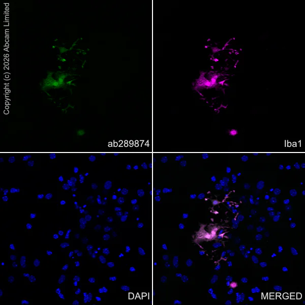
Immunocytochemistry/ Immunofluorescence - Anti-Iba1 antibody [EPR16588] - Goat IgG (Chimeric) - BSA and Azide free (AB289876)
This data was developed using the same antibody clone in a different buffer formulation containing PBS, BSA, glycerol, and sodium azide (ab289874).
Immunofluorescent analysis of 4% Paraformaldehyde-fixed, 0.1% TritonX-100 permeabilized mouse primary neural/glia cell cells labelling Iba1 with ab289874 at 1/1000 dilution , followed by ab150129 Donkey Anti-Goat IgG H&L (Alexa Fluor® 488) antibody at 1/500 dilution (Green).
Confocal image showing positive staining in mouse primary microglial cells (shown in green). The counterstain was observed in magenta. Nuclear DNA was labelled with DAPI (shown in blue). Image was taken with a confocal microscope(Leica-Microsystems, TCS SP8).
Confocal scanning Z step was set as 0.3 µm followed by image processing with maximum Z projection.
Rabbit anti-Iba1 antibody was used to counterstain Iba1 at 1/1000 dilution (Magenta). The Nuclear counterstain was DAPI (Blue).
Secondary antibody only control : Secondary antibody is ab150129 Donkey Anti-Goat IgG H&L (Alexa Fluor® 488) at 1/500 dilution.

- IHC-P
Lab
Immunohistochemistry (Formalin/PFA-fixed paraffin-embedded sections) - Anti-Iba1 antibody [EPR16588] - Goat IgG (Chimeric) - BSA and Azide free (AB289876)
This data was developed using ab289874, the same antibody clone in a different buffer formulation.
Immunohistochemical analysis of paraffin-embedded Rat liver tissue labeling Iba1 with ab289874 at 1/1000 dilution, followed by a ready to use LeicaDS9800 (Bond™ Polymer Refine Detection).
Positive staining on Kupffer cells in the rat liver. The section was incubated with ab289874 for 30 mins at room temperature and followed by specific anti-goat IgG antibody (ab97101) for 8 mins. The immunostaining was performed on a Leica Biosystems BOND®RX instrument.
Counterstained with Hematoxylin.
Secondary antibody only control : Secondary antibody is a ready to use LeicaDS9800 (Bond™ Polymer Refine Detection).
Heat mediated antigen retrieval with Tris-EDTA buffer (pH 9.0, epitope retrieval solution2) for 20 mins.
- ICC/IF
Lab
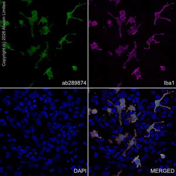
Immunocytochemistry/ Immunofluorescence - Anti-Iba1 antibody [EPR16588] - Goat IgG (Chimeric) - BSA and Azide free (AB289876)
This data was developed using the same antibody clone in a different buffer formulation containing PBS, BSA, glycerol, and sodium azide (ab289874).
Immunofluorescent analysis of 4% Paraformaldehyde-fixed, 0.1% TritonX-100 permeabilized rat primary neural/glia cell cells labelling Iba1 with ab289874 at 1/1000 dilution, followed by ab150129 Donkey Anti-Goat IgG H&L (Alexa Fluor® 488) antibody at 1/500 dilution (Green).
Confocal image showing positive staining in rat primary microglial cells (shown in green). The counterstain was observed in magenta. Nuclear DNA was labelled with DAPI (shown in blue). Image was taken with a confocal microscope(Leica-Microsystems, TCS SP8).
Confocal scanning Z step was set as 0.3 µm followed by image processing with maximum Z projection.
Rabbit anti-Iba1 antibody was used to counterstain Iba1 at 1/1000 dilution (Magenta). The Nuclear counterstain was DAPI (Blue).
Secondary antibody only control : Secondary antibody is ab150129 Donkey Anti-Goat IgG H&L (Alexa Fluor® 488) at 1/500 dilution.

- IHC-P
Lab
Immunohistochemistry (Formalin/PFA-fixed paraffin-embedded sections) - Anti-Iba1 antibody [EPR16588] - Goat IgG (Chimeric) - BSA and Azide free (AB289876)
This data was developed using ab289874, the same antibody clone in a different buffer formulation.
Immunohistochemical analysis of paraffin-embedded Mouse liver tissue labeling Iba1 with ab289874 at 1/1000 dilution, followed by a ready to use LeicaDS9800 (Bond™ Polymer Refine Detection).
Positive staining on Kupffer cells in the mouse liver.
The section was incubated with ab289874 for 30 mins at room temperature and followed by specific anti-goat IgG antibody (ab97101) for 8 mins. The immunostaining was performed on a Leica Biosystems BOND® RX instrument.
Counterstained with Hematoxylin.
Secondary antibody only control : Secondary antibody is a ready to use LeicaDS9800 (Bond™ Polymer Refine Detection).
Heat mediated antigen retrieval with Tris-EDTA buffer (pH 9.0, epitope retrieval solution2) for 20 mins.
관련 conjugated 항체와 다양한 조성의 항체 (5)
-
Anti-Iba1 antibody [EPR16588] - Microglia marker - Rat IgG2a (Chimeric)
-
Anti-Iba1 antibody [EPR16588] - Rat IgG2a (Chimeric) - BSA and Azide free
-
Anti-Iba1 antibody [EPR16588] - Microglia marker - Goat IgG (Chimeric)
-
Anti-Iba1 antibody [EPR16588] - Microglia marker – Chicken IgY (Chimeric)
-
Anti-Iba1 antibody [EPR16588] – Chicken IgY (Chimeric) – BSA and Azide Free
보완 제품


Primary Antibodies
AB278054
Anti-GFAP antibody [RM1003] - Astrocyte Marker
BOND RX™ Validated|20ul selling size|RabMAb|Recombinant

- 0
- 0
Primary Antibodies
AB177487
Anti-NeuN antibody [EPR12763] - Neuronal Marker
BOND RX™ Validated|RabMAb|Recombinant|Lab Essentials|20ul selling size|Advanced Validation

- 5
- 76
Primary Antibodies
AB183734
RabMAb|Recombinant|Lab Essentials|20ul selling size

- 5
- 24
Reactivity 정보
제품 세부 정보
ab289876 is the carrier-free version of ab289874.
This goat monoclonal chimeric antibody has been engineered from a RabMAb parent antibody (ab178846). By necessity, some rabbit sequence is retained as part of the variable domain. When multiplexing with other rabbit-derived antibodies, using cross absorbed FC-reactive secondary antibodies are recommended.
What are the advantages of a recombinant monoclonal antibody?
This product is a recombinant monoclonal antibody, which offers several advantages including:
- - High batch-to-batch consistency and reproducibility
- - Improved sensitivity and specificity
- - Long-term security of supply
- - Animal-free batch production
For more information, read more on recombinant antibodies.
Conjugation ready
Our carrier-free antibodies are typically supplied in a PBS-only formulation, purified and free of BSA, sodium azide and glycerol. This conjugation-ready format is designed for use with fluorochromes, metal isotopes, oligonucleotides, and enzymes, which makes them ideal for antibody labelling, functional and cell-based assays, flow-based assays (e.g. mass cytometry) and Multiplex Imaging applications.
Use our conjugation kits for antibody conjugates that are ready-to-use in as little as 20 minutes with 1 minute hands-on-time and 100% antibody recovery: available for fluorescent dyes, HRP, biotin and gold.
Compatibility
This product is compatible with the Maxpar® Antibody Labeling Kit from Fluidigm, without the need for antibody preparation. Maxpar® is a trademark of Fluidigm Canada Inc.
특성 및 보관 정보
제형
Purification 테크닉
보관 버퍼
배송 시 보관 조건
적절한 단기 보관 조건
적절한 장기 보관 조건
보관 정보
추가 정보
This supplementary information is collated from multiple sources and compiled automatically.
Biological function summary
Iba1 modulates actin bundling facilitating changes in microglial morphology and motility. It does not form part of a multiprotein complex but is significantly involved in cytoskeletal dynamics. This protein's expression increases during brain injury indicating its role in response to neural trauma. Iba1 has calcium-binding properties which suggest that it might interact with proteins involved in calcium signaling pathways.
Pathways
Calcium signaling and actin cytoskeletal rearrangement are significant biological processes that involve Iba1. Within these pathways Iba1 interacts closely with other actin-binding proteins that affect microglial movement and morphology. Such interactions illustrate how Iba1 helps microglia navigate and respond to changes within the brain environment.
제품 프로토콜
- Visit the General protocols
- Visit the Troubleshooting
타겟 정보
대체 명칭 보기
Product promise
Please note: All products are 'FOR RESEARCH USE ONLY. NOT FOR USE IN DIAGNOSTIC OR THERAPEUTIC PROCEDURES'.
For licensing inquiries, please contact partnerships@abcam.com