Anti-IL-6- antibody [EPRNGS_IL-6-20] BSA and Azide free, low endotoxin
- Recombinant
- RabMAb
- 각 태그의 뜻
Be the first to review this product! 리뷰 제출
|
(0 출판물)
- FuncS (Neut/Block)
Supplier Data
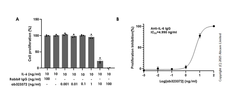
Functional Studies (Neut/Block) - Anti-IL-6- antibody [EPRNGS_IL-6-20] BSA and Azide free, low endotoxin (AB323372)
Cell proliferation of TF-1 (human erythroleukemia erythroblast) cells treated with IL-6 (ab259381) and anti-IL-6 antibody (ab323372) or isotype IgG (ab210849).
Cell proliferation (%) (A) confirms that the anti-IL-6 antibody (ab323372) neutralizes IL-6-mediated cell proliferation. The IC50 as determined by the dose-dependent inhibition of TF-1 cells proliferation is 4.990 ng/ml (B).
TF-1 cells were seeded at 5 × 10⁴ cells per well in a 96-well plate and cultured in medium containing 5% FBS for 48 h. Proliferation was analysed by luminescent assay and recorded using a Microplate Reader at 560nm emission.
- Flow Cyt (Intra)
Supplier Data
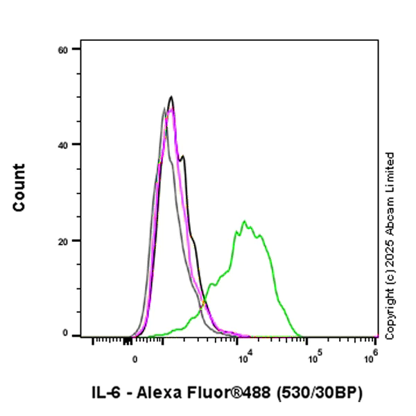
Flow Cytometry (Intracellular) - Anti-IL-6- antibody [EPRNGS_IL-6-20] BSA and Azide free, low endotoxin (AB323372)
Flow cytometric analysis of 4% paraformaldehyde fixed 90% methanol permeabilized HUVEC (human umbilical vein endothelial cell) treated with 0.5ug/mL LPS for 24 hours and 300ng/mL BFA for 20 hours (Green) / Untreated HUVEC (Magenta) cells labeling IL-6 with ab323372 at 1/1000 dilution (0.1 ug) / Magenta and Green compared with a Rabbit monoclonal IgG (ab172730) (Black) and (Grey) isotype control and an unlabeled control (cells without incubation with primary antibody and secondary antibody).
Goat Anti-Rabbit IgG (Alexa Fluor® 488, ab150081) at 1/2000 dilution was used as the secondary antibody.
- ICC/IF
Supplier Data
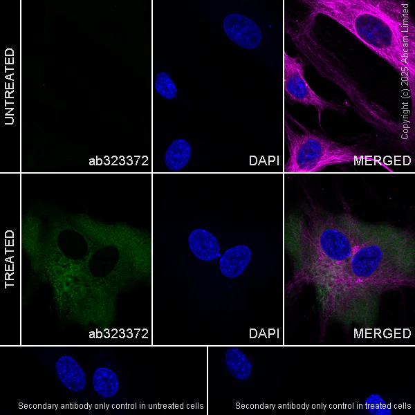
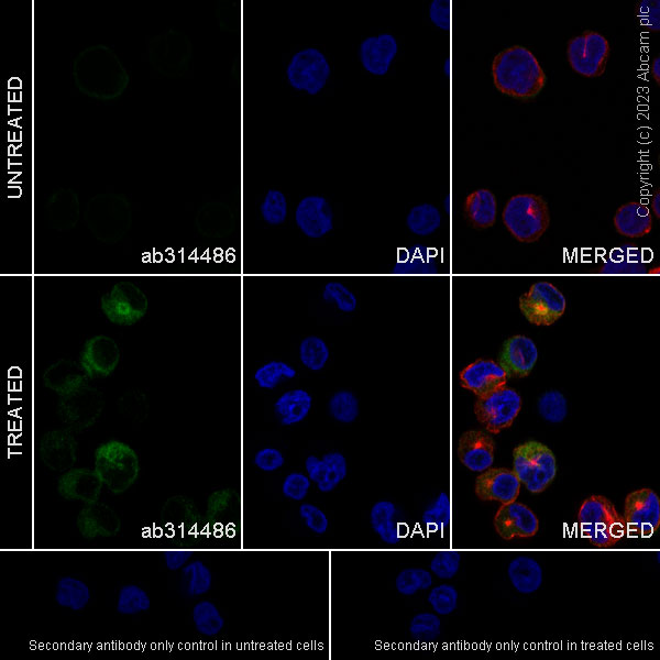
Immunocytochemistry/ Immunofluorescence - Anti-IL-6- antibody [EPRNGS_IL-6-20] BSA and Azide free, low endotoxin (AB323372)
Immunofluorescent analysis of 4% Paraformaldehyde-fixed, 0.1% TritonX-100 permeabilized HUVEC (human umbilical vein endothelial cell) cells labelling IL-6 with ab323372 at 1/100 (10 ug/ml) dilution, followed by ab150081 Goat Anti-Rabbit IgG H&L (Alexa Fluor® 488) preadsorbed antibody at 1/1000 2ug/ml dilution (Green).
Confocal image showing increased cytoplasmic staining in HUVEC cells treated with lipopolysaccharide (0.5 ug/ml) for 24 h, then Brefeldin A (300ng/ml) for another 20 h (shown in green). The counterstain was observed in magenta. Nuclear DNA was labelled with DAPI (shown in blue). Image was taken with a confocal microscope (Leica-Microsystems, TCS SP8).
ab195889 Anti-alpha Tubulin mouse monoclonal antibody - Microtubule Marker (Alexa Fluor® 594) was used to counterstain tubulin at 1/200 2.5ug/ml dilution (Magenta). The Nuclear counterstain was DAPI (Blue).
Secondary antibody only control : Secondary antibody is ab150081 Goat Anti-Rabbit IgG H&L (Alexa Fluor® 488) preadsorbed at 1/1000 2ug/ml dilution.
- IP
Supplier Data
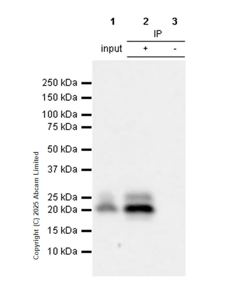
Immunoprecipitation - Anti-IL-6- antibody [EPRNGS_IL-6-20] BSA and Azide free, low endotoxin (AB323372)
IL-6 was immunoprecipitated from 0.35 mg HUVEC (human umbilical vein endothelial cell) treated with 0.5µg/ml LPS for 24h, 300ng/ml BFA was then added for additional 20h, whole cell lysate with ab323372 at 1/30 dilution (2ug in 0.35mg lysates). Western blot was performed on the immunoprecipitate using ab323372 at 1/1000 dilution. VeriBlot for IP secondary antibody(HRP)(ab131366) was used at 1/5000 dilution.
Lane 1 : HUVEC (human umbilical vein endothelial cell) treated with 0.5µg/ml LPS for 24h, 300ng/ml BFA was then added for additional 20h, whole cell lysate
Lane 2 : ab323372 IP in HUVEC (human umbilical vein endothelial cell) treated with 0.5µg/ml LPS for 24h, 300ng/ml BFA was then added for additional 20h, whole cell lysate
Lane 3 : Rabbit monoclonal IgG (ab172730) instead of ab323372 in HUVEC treated with 0.5µg/ml LPS for 24h, 300ng/ml BFA was then added for additional 20h, whole cell lysate
Blocking and dilution buffer and concentration : 5% NFDM/TBST
All lanes:
Immunoprecipitation - Anti-IL-6- antibody [EPRNGS_IL-6-20] BSA and Azide free, low endotoxin (ab323372) at 1/30 dilution
All lanes:
HUVEC (human umbilical vein endothelial cell) treated with 0.5µg/ml LPS for 24h, 300ng/ml BFA was then added for additional 20h, whole cell lysate
Secondary
All lanes:
Immunoprecipitation - VeriBlot for IP Detection Reagent (HRP) (<a href='/ko/products/reagents/veriblot-for-ip-detection-reagent-hrp-ab131366'>ab131366</a>) at 1/5000 dilution
false
Exposure time: 24s
- WB
Supplier Data
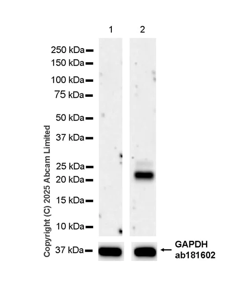
Western blot - Anti-IL-6- antibody [EPRNGS_IL-6-20] BSA and Azide free, low endotoxin (AB323372)
Blocking and diluting buffer and concentration : 5% NFDM/TBST.
The expression of IL-6 is upregulated in response to LPS and BFA treatment.
In Western blot, Anti-GAPDH antibody [EPR16891] - Loading Control (ab181602) staining at 1/200000 dilution.
All lanes:
Western blot - Anti-IL-6- antibody [EPRNGS_IL-6-20] BSA and Azide free, low endotoxin (ab323372) at 1/1000 dilution
Lane 1:
Untreated HUVEC (human umbilical vein endothelial cell) whole cell lysate at 20 µg
Lane 2:
HUVEC (human umbilical vein endothelial cell) treated with 0.5 ug/ml LPS for 24h, 300ng/ml BFA was then added for additional 20h, whole cell lysate at 20 µg
Secondary
All lanes:
Western blot - Goat Anti-Rabbit IgG H&L (HRP) (<a href='/ko/products/secondary-antibodies/goat-rabbit-igg-h-l-hrp-ab97051'>ab97051</a>) at 1/100000 dilution
Observed band size: 23-28 kDa,36 kDa
false
Exposure time: 180s
- I-ELISA
Supplier Data
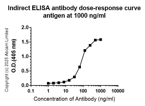
Indirect ELISA - Anti-IL-6- antibody [EPRNGS_IL-6-20] BSA and Azide free, low endotoxin (AB323372)
Indirect ELISA analysis of ab323372 at 1000-0 ng/ml. The Secondary antibody used was Alkaline Phosphatase-conjugated AffiniPure Goat Anti-Rabbit IgG (H+L) at 1 : 2500 dilution dilution.
Antigen : Human IL-6 protein.
Antigen concentration : 1000 ng/ml
보완 제품


Primary Antibodies
AB289967
Anti-IL-8 antibody [EPR26511-74]
20ul selling size|RabMAb|Recombinant|KO Validated

- 0
- 0
Primary Antibodies
AB307164
Anti-TNF alpha antibody [RM1005]
BOND RX™ Validated|Recombinant|Lab Essentials|20ul selling size|RabMAb

- 0
- 0
Primary Antibodies
AB314487
Anti-IL-23 antibody [EPR26364-40] - BSA and Azide free
RabMAb|Recombinant
- 0
- 0
Primary Antibodies
AB283575
BOND RX™ Validated|20ul selling size|RabMAb|Recombinant|KO Validated|Trial Size

- 0
- 0
Reactivity 정보
제품 세부 정보
Patented technology
Our RabMAb® technology is a patented hybridoma-based technology for making rabbit monoclonal antibodies. For details on our patents, please refer to RabMAb® patents.
What are the advantages of a recombinant monoclonal antibody?
This product is a recombinant monoclonal antibody, which offers several advantages including:
- - High batch-to-batch consistency and reproducibility
- - Improved sensitivity and specificity
- - Long-term security of supply
- - Animal-free batch production
For more information, read more on recombinant antibodies.
Compatibility
This product is compatible with the Maxpar® Antibody Labeling Kit from Fluidigm, without the need for antibody preparation. Maxpar® is a trademark of Fluidigm Canada Inc.
Conjugation ready
Our carrier-free antibodies are typically supplied in a PBS-only formulation, purified and free of BSA, sodium azide and glycerol. This conjugation-ready format is designed for use with fluorochromes, metal isotopes, oligonucleotides, and enzymes, which makes them ideal for antibody labelling, functional and cell-based assays, flow-based assays (e.g. mass cytometry) and Multiplex Imaging applications.
Use our conjugation kits for antibody conjugates that are ready-to-use in as little as 20 minutes with 1 minute hands-on-time and 100% antibody recovery: available for fluorescent dyes, HRP, biotin and gold.
특성 및 보관 정보
제형
Purification 테크닉
보관 버퍼
배송 시 보관 조건
적절한 단기 보관 조건
적절한 장기 보관 조건
제품 프로토콜
- Visit the General protocols
- Visit the Troubleshooting
타겟 정보
대체 명칭 보기
Product promise
Please note: All products are 'FOR RESEARCH USE ONLY. NOT FOR USE IN DIAGNOSTIC OR THERAPEUTIC PROCEDURES'.
For licensing inquiries, please contact partnerships@abcam.com