Anti-Oxytocin Receptor antibody [EPR24409-146]- BSA and Azide free
- RabMAb
- Recombinant
- 각 태그의 뜻
3
(2 리뷰들)
|
(0 출판물)
- ICC/IF
Supplier Data
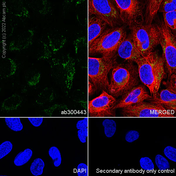
Immunocytochemistry/ Immunofluorescence - Anti-Oxytocin Receptor antibody [EPR24409-146]- BSA and Azide free (AB300444)
This data was developed using ab300443, the same antibody clone in a different buffer formulation.Immunofluorescent analysis of 4% Paraformaldehyde-fixed, 0.1% TritonX-100 permeabilized U-2 OS (human bone osteosarcoma epithelial cell) cells labelling Oxytocin Receptor with ab300443 at 1/50 (9.8 ug/ml) dilution, followed by ab150081 Goat Anti-Rabbit IgG H&L (Alexa Fluor® 488) preadsorbed antibody at 1/1000 2 ug/ml dilution (Green). Confocal image showing cytoplasmic staining in U-2 OS cell line is observed. ab195889 Anti-alpha Tubulin mouse monoclonal antibody - Microtubule Marker (Alexa Fluor® 594) was used to counterstain tubulin at 1/200 2.5 ug/ml dilution (Red). The Nuclear counterstain was DAPI (Blue). Secondary antibody only control : Secondary antibody is ab150081 Goat Anti-Rabbit IgG H&L (Alexa Fluor® 488) preadsorbed at 1/1000 2 ug/ml dilution.
- ICC/IF
Supplier Data
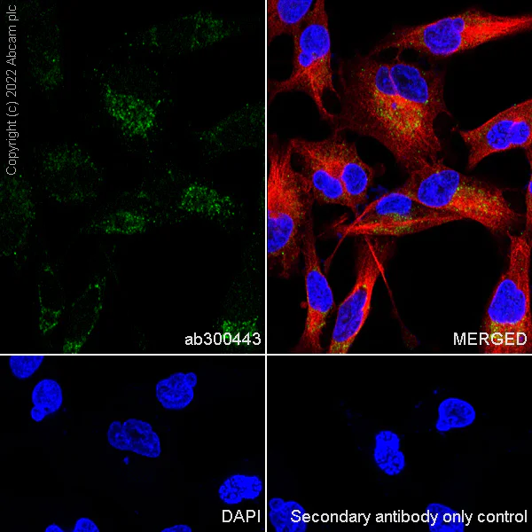
Immunocytochemistry/ Immunofluorescence - Anti-Oxytocin Receptor antibody [EPR24409-146]- BSA and Azide free (AB300444)
This data was developed using ab300443, the same antibody clone in a different buffer formulation.Immunofluorescent analysis of 4% Paraformaldehyde-fixed, 0.1% TritonX-100 permeabilized Rat-1(rat embryonic fibroblast) cells labelling Oxytocin Receptor with ab300443 at 1/50 (9.8 ug/ml) dilution, followed by ab150081 Goat Anti-Rabbit IgG H&L (Alexa Fluor® 488) preadsorbed antibody at 1/1000 2 ug/ml dilution (Green). Confocal image showing cytoplasmic staining in Rat-1 cell line is observed. ab195889 Anti-alpha Tubulin mouse monoclonal antibody - Microtubule Marker (Alexa Fluor® 594) was used to counterstain tubulin at 1/200 2.5 ug/ml dilution (Red). The Nuclear counterstain was DAPI (Blue). Secondary antibody only control : Secondary antibody is ab150081 Goat Anti-Rabbit IgG H&L (Alexa Fluor® 488) preadsorbed at 1/1000 2 ug/ml dilution.
- WB
Lab
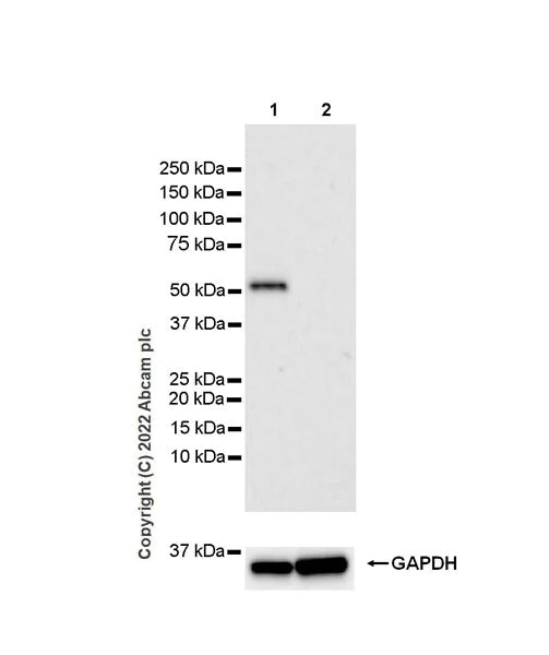
Western blot - Anti-Oxytocin Receptor antibody [EPR24409-146]- BSA and Azide free (AB300444)
This data was developed using 300443, the same antibody clone in a different buffer formulation.
Blocking and diluting buffer and concentration :
The expression profile/ molecular weight observed is consistent with what has been described in the literature (PMID : 11467983).
Negative control : skeletal muscle (PMID : 11467983).
All lanes:
Western blot - Anti-Oxytocin Receptor antibody [EPR24409-146] (<a href='/ko/products/primary-antibodies/oxytocin-receptor-antibody-epr24409-146-ab300443'>ab300443</a>) at 1/1000 dilution
Lane 1:
Human testis tissue lysate at 20 µg
Lane 2:
Human skeletal muscle tissue lysate at 20 µg
Secondary
All lanes:
Goat Anti-Rabbit IgG (HRP) with minimal cross-reactivity with human IgG at 1/2000 dilution
Predicted band size: 43 kDa
Observed band size: 55 kDa
false
Exposure time: 3min
- WB
Lab
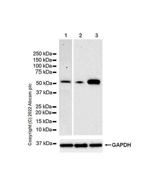
Western blot - Anti-Oxytocin Receptor antibody [EPR24409-146]- BSA and Azide free (AB300444)
This data was developed using 300443, the same antibody clone in a different buffer formulation.
Blocking and diluting buffer and concentration : 5% NFDM/TBST.
The expression profile/ molecular weight observed is consistent with what has been described in the literature (PMID : 28382233).
The blot of lane 1 was developed using a high sensitivity ECL substrate.
All lanes:
Western blot - Anti-Oxytocin Receptor antibody [EPR24409-146] (<a href='/ko/products/primary-antibodies/oxytocin-receptor-antibody-epr24409-146-ab300443'>ab300443</a>) at 1/1000 dilution
Lane 1:
U-2 OS (human bone osteosarcoma epithelial cell), whole cell lysate at 20 µg
Lane 2:
Untreated C2C12 (mouse myoblasts myoblast), whole cell lysate at 20 µg
Lane 3:
C2C12 differentiation for 7 days, whole cell lysate at 20 µg
Secondary
All lanes:
Western blot - Goat Anti-Rabbit IgG H&L (HRP) (<a href='/ko/products/secondary-antibodies/goat-rabbit-igg-h-l-hrp-ab97051'>ab97051</a>) at 1/100000 dilution
Predicted band size: 42 kDa
Observed band size: 55 kDa
false
Exposure time: 3min
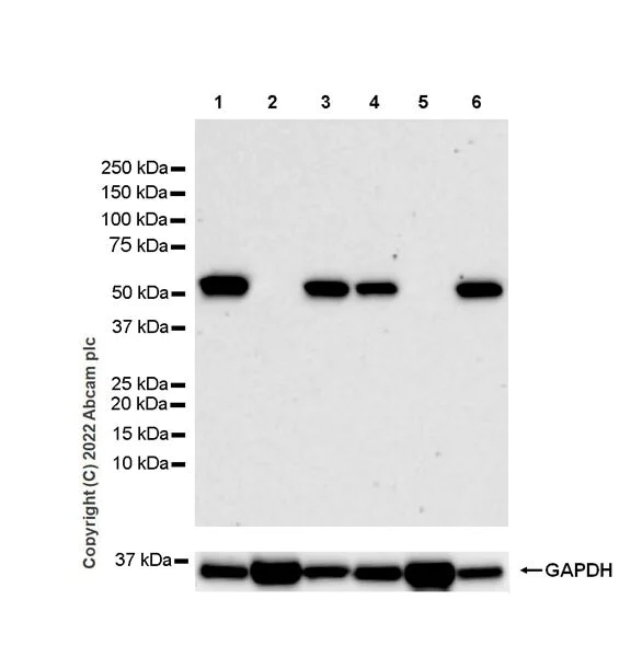
- WB
Lab
Western blot - Anti-Oxytocin Receptor antibody [EPR24409-146]- BSA and Azide free (AB300444)
This data was developed using 300443, the same antibody clone in a different buffer formulation.
Blocking and diluting buffer and concentration : The expression profile/ molecular weight observed is consistent with what has been described in the literature (PMID : 11467983).
Negative control : skeletal muscle (PMID : 11467983).
All lanes:
Western blot - Anti-Oxytocin Receptor antibody [EPR24409-146] (<a href='/ko/products/primary-antibodies/oxytocin-receptor-antibody-epr24409-146-ab300443'>ab300443</a>) at 1/1000 dilution
Lane 1:
Rat uterus tissue lysate at 20 µg
Lane 2:
Rat skeletal muscle tissue lysate at 20 µg
Lane 3:
Rat testis tissue lysate at 20 µg
Lane 4:
Mouse uterus tissue lysate at 20 µg
Lane 5:
Mouse skeletal muscle tissue lysate at 20 µg
Lane 6:
Mouse testis tissue lysate at 20 µg
Secondary
All lanes:
Western blot - Goat Anti-Rabbit IgG H&L (HRP) (<a href='/ko/products/secondary-antibodies/goat-rabbit-igg-h-l-hrp-ab97051'>ab97051</a>) at 1/100000 dilution
Predicted band size: 42 kDa
Observed band size: 55 kDa
false
Exposure time: 3min
관련 conjugated 항체와 다양한 조성의 항체 (1)
-
Anti-Oxytocin Receptor antibody [EPR24409-146]
보완 제품


Primary Antibodies
AB212193
Anti-Oxytocin-neurophysin 1 antibody [EPR20973]
20ul selling size|RabMAb|Recombinant

- 4
- 1
Primary Antibodies
AB302667
BOND RX™ Validated|Recombinant|Lab Essentials|KO Validated

- 0
- 0
Primary Antibodies
AB183734
RabMAb|Recombinant|Lab Essentials|20ul selling size

- 5
- 24
Primary Antibodies
AB124964
Anti-alpha smooth muscle Actin antibody [EPR5368]
BOND RX™ Validated|RabMAb|Recombinant|KO Validated|20ul selling size

- 5
- 19
Reactivity 정보
제품 세부 정보
Patented technology
Our RabMAb® technology is a patented hybridoma-based technology for making rabbit monoclonal antibodies. For details on our patents, please refer to RabMAb® patents.
What are the advantages of a recombinant monoclonal antibody?
This product is a recombinant monoclonal antibody, which offers several advantages including:
- - High batch-to-batch consistency and reproducibility
- - Improved sensitivity and specificity
- - Long-term security of supply
- - Animal-free batch production
For more information, read more on recombinant antibodies.
Conjugation ready
Our carrier-free antibodies are typically supplied in a PBS-only formulation, purified and free of BSA, sodium azide and glycerol. This conjugation-ready format is designed for use with fluorochromes, metal isotopes, oligonucleotides, and enzymes, which makes them ideal for antibody labelling, functional and cell-based assays, flow-based assays (e.g. mass cytometry) and Multiplex Imaging applications.
Use our conjugation kits for antibody conjugates that are ready-to-use in as little as 20 minutes with 1 minute hands-on-time and 100% antibody recovery: available for fluorescent dyes, HRP, biotin and gold.
Compatibility
This product is compatible with the Maxpar® Antibody Labeling Kit from Fluidigm, without the need for antibody preparation. Maxpar® is a trademark of Fluidigm Canada Inc.
특성 및 보관 정보
제형
Purification 테크닉
보관 버퍼
배송 시 보관 조건
적절한 단기 보관 조건
적절한 장기 보관 조건
보관 정보
추가 정보
This supplementary information is collated from multiple sources and compiled automatically.
Biological function summary
The oxytocin receptor exhibits involvement in many reproductive and social behaviors. Despite not functioning as part of a larger complex OTR activation facilitates processes such as uterine contraction milk ejection and social bonding. Activation of this receptor influences neuronal circuits in the brain supporting behaviors linked to emotion and social memory which are fundamental to maternal behaviors and pair bonding.
Pathways
The oxytocin receptor participates in critical signaling cascades including the phosphatidylinositol-calcium second messenger system. Through this pathway the receptor activates protein kinase C and increases intracellular calcium levels. This pathway often involves interaction with other proteins such as vasopressin receptors which have overlapping functions in fluid balance and cardiovascular regulation.
제품 프로토콜
- Visit the General protocols
- Visit the Troubleshooting
타겟 정보
대체 명칭 보기
Product promise
Please note: All products are 'FOR RESEARCH USE ONLY. NOT FOR USE IN DIAGNOSTIC OR THERAPEUTIC PROCEDURES'.
For licensing inquiries, please contact partnerships@abcam.com