Anti-PAR1/Thrombin Receptor antibody [EPR28398-8]
- KD Validated
- 20ul selling size
- BOND RX™ Validated
- Advanced Validation
- RabMAb
- Recombinant
- 각 태그의 뜻
Be the first to review this product! 리뷰 제출
|
(0 출판물)

- mIHC
Lab
Multiplex immunohistochemistry - Anti-PAR1/Thrombin Receptor antibody [EPR28398-8] (AB322457)
Multiplex immunohistochemistry analysis of formalin/PFA-fixed paraffin-embedded mouse liver tissue staining ALDH1L1 with ab307696 at a 1/5000 dilution, ab322457 anti-PAR1 used at 1/500 dilution and ab315474 anti-ABCB11 used at a 1/2000 dilution.
Panel A : merged staining of anti-ALDH1L1 (green; Opal™520), anti-PAR1 (magenta; Opal™690) and anti-ABCB11 (gray; Opal™570) on mouse liver.
Panel B : anti-ALDH1L1 staining hepatocytes in mouse liver.
Panel C : anti-PAR1 staining endothelium in mouse liver.
Panel D : anti-ABCB11 staining bile canaliculi in mouse liver.
Nuclear DNA was labeled with DAPI (shown in blue).
The section was incubated in three rounds of staining : in the order of ab307696, ab322457 and ab315474 for 30 mins at room temperature. Each round was followed by a separate fluorescent tyramide signal amplification system.
Heat mediated antigen retrieval with Tris-EDTA buffer (pH 9.0, epitope retrieval solution2) for 20 mins.

- IHC-P
Supplier Data
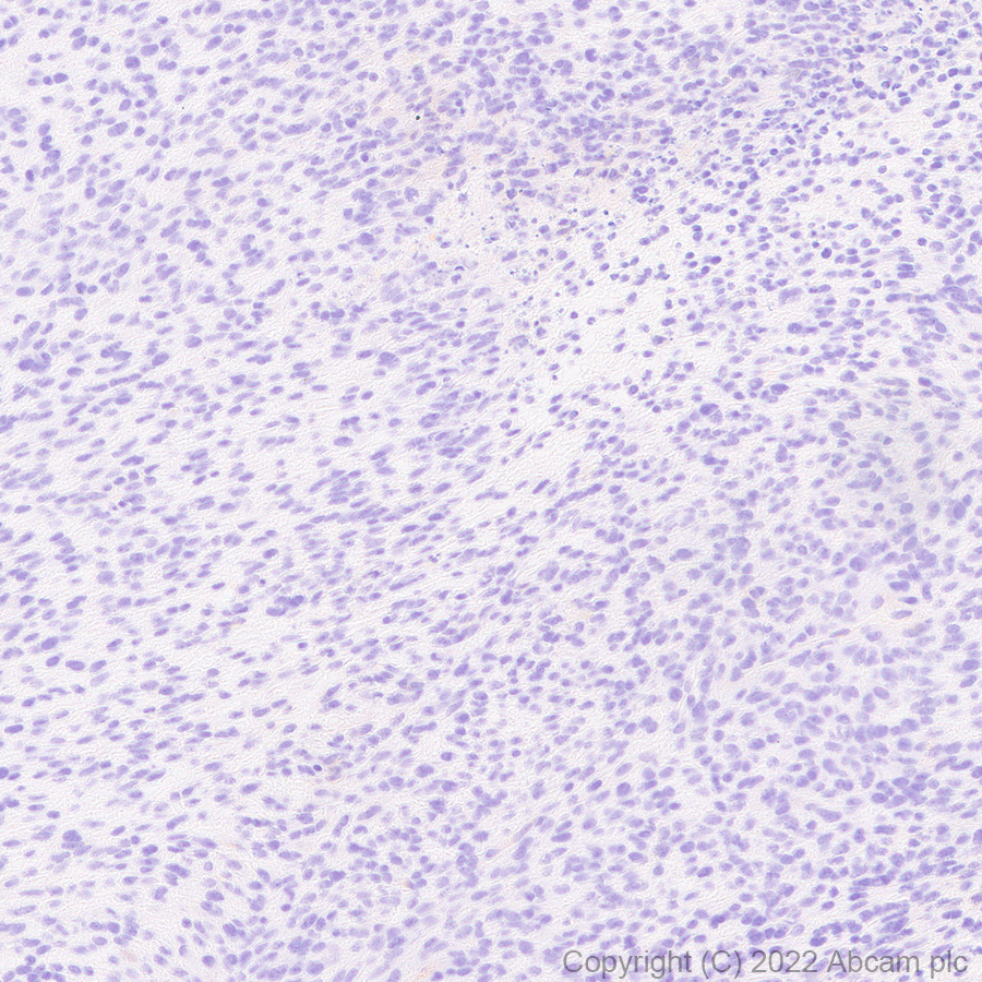
Immunohistochemistry (Formalin/PFA-fixed paraffin-embedded sections) - Anti-PAR1/Thrombin Receptor antibody [EPR28398-8] (AB322457)
Immunohistochemical analysis of paraffin-embedded Mouse spleen tissue labeling PAR1/Thrombin Receptor with ab322457 at 1/500 (1.054 ug/ml) dilution, followed by a ready to use LeicaDS9800 (Bond™ Polymer Refine Detection).
Positive staining on mouse spleen.
The section was incubated with ab322457 for 30 mins at room temperature.
The immunostaining was performed on a Leica Biosystems BOND® RX instrument
Counterstained with Hematoxylin.
Secondary antibody only control : Secondary antibody is a ready to use LeicaDS9800 (Bond™ Polymer Refine Detection).
Heat mediated antigen retrieval was performed with Tris-EDTA buffer (pH 9.0, Epitope Retrieval Solution2) for 20 mins

- IHC
Lab
Immunohistochemistry - Anti-PAR1/Thrombin Receptor antibody [EPR28398-8] (AB322457)
Multiplex immunohistochemistry analysis of formalin/PFA-fixed paraffin-embedded mouse kidney tissue staining PAR1/Thrombin Receptor with ab322457 at a 1/500 dilution, ab306558 anti-SGLT2 used at 1/5000 dilution and ab212197 anti-Renin used at a 1/4000 dilution.
Panel A : merged staining of anti-PAR1/Thrombin Receptor (green; Opal™520), anti-SGLT2 (magenta; Opal™690) and anti-Renin (gray; Opal™570) on mouse kidney.
Panel B : anti-PAR1/Thrombin Receptor staining endothelium in mouse kidney.
Panel C : anti-SGLT2 staining the brush border of proximal tubules in mouse kidney.
Panel D : anti-Renin staining juxtaglomerular cells in mouse kidney.
Nuclear DNA was labeled with DAPI (shown in blue).
The section was incubated in three rounds of staining : in the order of ab322457, ab306558 and ab212197 for 30 mins at room temperature. Each round was followed by a separate fluorescent tyramide signal amplification system.
The immunostaining was performed on a Leica Biosystems BOND® RX instrument with an Opal™ 4-color kit. Image acquisition was performed with Leica SP8 confocal microscope.
Heat mediated antigen retrieval with Tris-EDTA buffer (pH 9.0, epitope retrieval solution2) for 20 mins.

- IHC-P
Supplier Data
Immunohistochemistry (Formalin/PFA-fixed paraffin-embedded sections) - Anti-PAR1/Thrombin Receptor antibody [EPR28398-8] (AB322457)
Immunohistochemical analysis of paraffin-embedded Mouse lung tissue labeling PAR1/Thrombin Receptor with ab322457 at 1/500 (1.054 ug/ml) dilution, followed by a ready to use LeicaDS9800 (Bond™ Polymer Refine Detection).
Positive staining on mouse lung (PMID : 12548614).
The section was incubated with ab322457 for 30 mins at room temperature.
The immunostaining was performed on a Leica Biosystems BOND® RX instrument
Counterstained with Hematoxylin.
Secondary antibody only control : Secondary antibody is a ready to use LeicaDS9800 (Bond™ Polymer Refine Detection).
Heat mediated antigen retrieval was performed with Tris-EDTA buffer (pH 9.0, Epitope Retrieval Solution2) for 20 mins

- IHC-P
Supplier Data
Immunohistochemistry (Formalin/PFA-fixed paraffin-embedded sections) - Anti-PAR1/Thrombin Receptor antibody [EPR28398-8] (AB322457)
Immunohistochemical analysis of paraffin-embedded Mouse liver tissue labeling PAR1/Thrombin Receptor with ab322457 at 1/500 (1.054 ug/ml) dilution, followed by a ready to use LeicaDS9800 (Bond™ Polymer Refine Detection).
Positive staining on mouse liver.
The section was incubated with ab322457 for 30 mins at room temperature.
The immunostaining was performed on a Leica Biosystems BOND® RX instrument
Counterstained with Hematoxylin.
Secondary antibody only control : Secondary antibody is a ready to use LeicaDS9800 (Bond™ Polymer Refine Detection).
Heat mediated antigen retrieval was performed with Tris-EDTA buffer (pH 9.0, Epitope Retrieval Solution2) for 20 mins

- IHC-P
Supplier Data
Immunohistochemistry (Formalin/PFA-fixed paraffin-embedded sections) - Anti-PAR1/Thrombin Receptor antibody [EPR28398-8] (AB322457)
Immunohistochemical analysis of paraffin-embedded Mouse kidney tissue labeling PAR1/Thrombin Receptor with ab322457 at 1/500 (1.054 ug/ml) dilution, followed by a ready to use LeicaDS9800 (Bond™ Polymer Refine Detection).
Positive staining on mouse kidney.
The section was incubated with ab322457 for 30 mins at room temperature.
The immunostaining was performed on a Leica Biosystems BOND® RX instrument
Counterstained with Hematoxylin.
Secondary antibody only control : Secondary antibody is a ready to use LeicaDS9800 (Bond™ Polymer Refine Detection).
Heat mediated antigen retrieval was performed with Tris-EDTA buffer (pH 9.0, Epitope Retrieval Solution2) for 20 mins

- WB
Supplier Data
Western blot - Anti-PAR1/Thrombin Receptor antibody [EPR28398-8] (AB322457)
Blocking and diluting buffer and concentration : 5% NFDM/TBST.
PAR1 contains different glycosylation sites, the multiple bands may be due to glycosylation (PMID : 20368337).
This blot was developed using a high-sensitivity ECL substrate, allowing for the detection of proteins in the mid-femtogram range.
In Western blot, Anti-GAPDH antibody [EPR16891] - Loading Control (ab181602) staining at 1/200000 dilution.
All lanes:
Western blot - Anti-PAR1/Thrombin Receptor antibody [EPR28398-8] (ab322457) at 1/1000 dilution
Lane 1:
Neuro-2a (mouse neuroblastoma neuroblast) transfected with scrambled siRNA control whole cell lysate at 20 µg
Lane 2:
Neuro-2a transfected with siRNA specifically targeting PAR1 whole cell lysate at 20 µg
Secondary
All lanes:
Western blot - Goat Anti-Rabbit IgG H&L (HRP) (<a href='/ko/products/secondary-antibodies/goat-rabbit-igg-h-l-hrp-ab97051'>ab97051</a>) at 1/20000 dilution
Observed band size: 47-75 kDa,36 kDa
true
Exposure time: 81s

- WB
Supplier Data
Western blot - Anti-PAR1/Thrombin Receptor antibody [EPR28398-8] (AB322457)
Blocking and diluting buffer and concentration : 5% NFDM/TBST.
Low expression : RAW 264.7, HL-1.
PAR1 contains different glycosylation sites, the multiple bands may be due to glycosylation (PMID : 20368337).
In Western blot, Anti-GAPDH antibody [EPR16891] - Loading Control (ab181602) staining at 1/200000 dilution.
All lanes:
Western blot - Anti-PAR1/Thrombin Receptor antibody [EPR28398-8] (ab322457) at 1/1000 dilution
Lane 1:
Mouse dorsal galion tissue lysate at 20 µg
Lane 2:
Neuro-2a (mouse neuroblastoma neuroblast) whole cell lysate at 20 µg
Lane 3:
RAW 264.7 (mouse abelson murine leukemia virus-induced tumor macrophage) whole cell lysate at 20 µg
Lane 4:
HL-1 (mouse atrial muscle cell) whole cell lysate at 20 µg
Lane 5:
C8-D1A (mouse cerebellum astrocyte) whole cell lysate at 20 µg
Lane 6:
SW10 (mouse neuronal schwann cell) whole cell lysate at 20 µg
Lane 7:
NIH/3T3 (mouse embryonic fibroblast) whole cell lysate at 20 µg
Secondary
All lanes:
Western blot - Goat Anti-Rabbit IgG H&L (HRP) (<a href='/ko/products/secondary-antibodies/goat-rabbit-igg-h-l-hrp-ab97051'>ab97051</a>) at 1/20000 dilution
Observed band size: 48-75 kDa,36 kDa
false
Exposure time: 136s
관련 conjugated 항체와 다양한 조성의 항체 (1)
-
Anti-PAR1/Thrombin Receptor antibody [EPR28398-8] - BSA and Azide free
보완 제품

Primary Antibodies
AB76302
Anti-NF-kB p65 (phospho S536) antibody [EP2294Y]
20ul selling size|RabMAb|Recombinant

- 4
- 7
Primary Antibodies
AB32536
Anti-NF-kB p65 antibody [E379]
BOND RX™ Validated|20ul selling size|RabMAb|Recombinant|KO Validated

- 4
- 15

Primary Antibodies
AB302911
Anti-EPCR/CD201 antibody [EPR26267-44]
BOND RX™ Validated|20ul selling size|RabMAb|Recombinant
- 0
- 0
Reactivity 정보
제품 세부 정보
Patented technology
Our RabMAb® technology is a patented hybridoma-based technology for making rabbit monoclonal antibodies. For details on our patents, please refer to RabMAb® patents.
What are the advantages of a recombinant monoclonal antibody?
This product is a recombinant monoclonal antibody, which offers several advantages including:
- - High batch-to-batch consistency and reproducibility
- - Improved sensitivity and specificity
- - Long-term security of supply
- - Animal-free batch production
For more information, read more on recombinant antibodies.
특성 및 보관 정보
제형
Purification 테크닉
보관 버퍼
배송 시 보관 조건
적절한 단기 보관 기간
적절한 단기 보관 조건
적절한 장기 보관 조건
분주 정보
보관 정보
제품 프로토콜
- Visit the General protocols
- Visit the Troubleshooting
타겟 정보
대체 명칭 보기
Product promise
Please note: All products are 'FOR RESEARCH USE ONLY. NOT FOR USE IN DIAGNOSTIC OR THERAPEUTIC PROCEDURES'.
For licensing inquiries, please contact partnerships@abcam.com