Anti-Paxillin antibody [Y113] - BSA and Azide free
- BOND RX™ Validated
- RabMAb
- Recombinant
- KO Validated
- 각 태그의 뜻
Be the first to review this product! 리뷰 제출
|
(14 제품이 사용된 논문 )

- Flow Cyt (Intra)
Unknown
Flow Cytometry (Intracellular) - Anti-Paxillin antibody [Y113] - BSA and Azide free (AB216652)
Intracellular Flow Cytometry analysis of HeLa (human cervix adenocarcinoma) cells labeling with purified ab32084 at 1/100 dilution (10ug/ml) (Red). Cells were fixed with 4% paraformaldehyde and permeabilised with 90% methanol. A Goat anti rabbit IgG (Alexa Fluor® 488) (ab150077) (1/2000 dilution) was used as the secondary antibody. Rabbit monoclonal IgG (Black) (ab172730) was used as a isotype control.Cell without incubation with primary antibody and secondary antibody (Blue) were used as unlabeled control.
This data was developed using the same antibody clone in a different buffer formulation containing PBS, BSA, glycerol, and sodium azide (ab32084).

- IHC-P
Supplier Data
Immunohistochemistry (Formalin/PFA-fixed paraffin-embedded sections) - Anti-Paxillin antibody [Y113] - BSA and Azide free (AB216652)
Immunohistochemistry analysis of paraffin-embedded human breast carcinoma tissue sections labelling Paxillin with ab32084 at 1/1200 dilution. The section was incubated with ab32084 for 30 mins at room temperature. Rabbit specific IHC polymer detection kit HRP/DAB (ab209101) was used as the secondary antibody. Sections were counterstained with Hematoxylin. Antigen retrieval was heat mediated antigen retrieval with Citrate buffer (pH 6.0, epitope retrieval solution 1) for 20 mins.
Positive staining on human breast carcinoma tissue. The immunostaining was performed on a Leica Biosystems BOND® RX instrument.
This data was developed using ab32084, the same antibody clone in a different buffer formulation.
- ICC/IF
Supplier Data
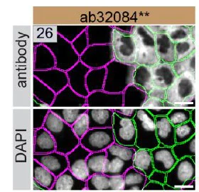
Immunocytochemistry/ Immunofluorescence - Anti-Paxillin antibody [Y113] - BSA and Azide free (AB216652)
This data was developed using ab32084, the same antibody clone in a different buffer formulation.
ab32084 was shown to react with PXN in wild-type A-431 cells in immunocytochemistry with loss of signal observed in PXN knockout cell line ab261892. Wild-type and knockout cells were mixed and pelleted at a 1 : 1 ratio on coverslips. The cells were fixed with 4% paraformaldehyde (15 min) then permeabilized with 0.1% Triton X-100 (10min) and then blocked with 1x PBS, 0.01% Triton X-100, 5% BSA, 5% NGS. The cells were then incubated with ab32084 at 1/150 dilution overnight at 4°C followed by a further incubation at room temperature for 1h with a goat anti-rabbit secondary antibody to (Alexa Fluor® 555) at 0.5 μg/ml. Acquisition of the green (wild-type), red (antibody staining) and far-red (knockout) channels was performed. Representative grayscale images of the red channel are shown. Wild-type and knockout cells are outlined with yellow and magenta dashed line, respectively. Schematic representation of the mosaic strategy used is shown on the bottom-right panel. Image was acquired with a Zeiss(LSM-880).
This data was provided by YCharOS Inc., an open science company with the mission of characterizing commercially available antibody reagents for all human proteins. Abcam and YCharOS are working together to help address the reproducibility crisis by enabling the life science community to better evaluate commercially available antibodies.

- IHC-P
Supplier Data
Immunohistochemistry (Formalin/PFA-fixed paraffin-embedded sections) - Anti-Paxillin antibody [Y113] - BSA and Azide free (AB216652)
Immunohistochemistry analysis of paraffin-embedded human cerebrum tissue sections labelling Paxillin with ab32084 at 1/1200 dilution. The section was incubated with ab32084 for 30 mins at room temperature. Rabbit specific IHC polymer detection kit HRP/DAB (ab209101) was used as the secondary antibody. Sections were counterstained with Hematoxylin. Antigen retrieval was heat mediated antigen retrieval with Citrate buffer (pH 6.0, epitope retrieval solution 1) for 20 mins.
Positive staining on human cerebrum tissue. The immunostaining was performed on a Leica Biosystems BOND® RX instrument.
This data was developed using ab32084, the same antibody clone in a different buffer formulation.

- ICC/IF
Supplier Data
Immunocytochemistry/ Immunofluorescence - Anti-Paxillin antibody [Y113] - BSA and Azide free (AB216652)
This data was developed using ab32084, the same antibody clone in a different buffer formulation.
Immunocytochemistry analysis of HeLa (Human cervix adenocarcinoma epithelial cell) cells labeling Paxillin with purified ab32084 at 1/50 dilution (2.88 μg/mL). Cells were fixed in 4% Paraformaldehyde and permeabilized with 0.1% tritonX-100. Cells were counterstained with ab195889 Anti-alpha Tubulin antibody [DM1A] - Microtubule Marker (Alexa Fluor® 594) 1/200 (2.5 μg/mL). Goat anti rabbit IgG (Alexa Fluor® 488, ab150077) was used as the secondary antibody at 1/1000 (2 μg/mL) dilution. DAPI (blue) was used as nuclear counterstain. PBS instead of the primary antibody was used as the secondary antibody only control.

- IP
Lab
Immunoprecipitation - Anti-Paxillin antibody [Y113] - BSA and Azide free (AB216652)
This data was developed using ab32084, the same antibody clone in a different buffer formulation.
Purified ab32084 at 1/20 dilution (0.7μg) immunoprecipitating Paxillin in HeLa whole cell lysate.
Lane 1 (input) : HeLa (Human cervix adenocarcinoma epithelial cell) whole cell lysate 10μg.
Lane 2 (+) : ab32084 + HeLa whole cell lysate.
Lane 3 (-) : Rabbit monoclonal IgG (ab172730) instead of ab32084 in HeLa whole cell lysate.
VeriBlot for IP Detection Reagent (HRP) (ab131366) (1/1000 dilution) was used for Western blotting.
Blocking Buffer and concentration : 5% NFDM/TBST.
Diluting buffer and concentration : 5% NFDM/TBST.
All lanes:
Immunoprecipitation - Anti-Paxillin antibody [Y113] (<a href='/ko/products/primary-antibodies/paxillin-antibody-y113-ab32084'>ab32084</a>)
Predicted band size: 65 kDa
Observed band size: 68 kDa
false
- IP
Supplier Data
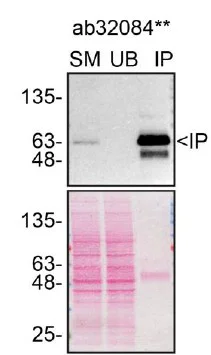
Immunoprecipitation - Anti-Paxillin antibody [Y113] - BSA and Azide free (AB216652)
This data was developed using ab32084, the same antibody clone in a different buffer formulation.
Immunoprecipitation of PXN in A-431 cells. Lysates were prepared and immunoprecipitation was performed using 13.6 μg of ab32084 pre-coupled to Protein A beads. Samples were then washed and processed for western blot.
This data was provided by YCharOS Inc., an open science company with the mission of characterizing commercially available antibody reagents for all human proteins. Abcam and YCharOS are working together to help address the reproducibility crisis by enabling the life science community to better evaluate commercially available antibodies.
All lanes:
Immunoprecipitation - Anti-Paxillin antibody [Y113] (<a href='/ko/products/primary-antibodies/paxillin-antibody-y113-ab32084'>ab32084</a>) at 13.6 µg
All lanes:
A-431 cell Lysates
Observed band size: 64 kDa
false
- WB
Supplier Data
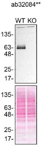
Western blot - Anti-Paxillin antibody [Y113] - BSA and Azide free (AB216652)
This data was developed using ab32084, the same antibody clone in a different buffer formulation.
ab32084 was shown to react with PXN in wild-type A-431 cells in Western blot with loss of signal observed in PXN knockout cell line ab261892. Wild-type A-431 and PXN knockout cell lysates were subjected to SDS-PAGE. Membranes were blocked in 5% milk in TBST for 1 hr before incubation with ab32084 overnight at 4 °C at a 1/1000 dilution. Blots were incubated with secondary antibodies at 0.2 µg/mL before imaging.
This data was provided by YCharOS Inc., an open science company with the mission of characterizing commercially available antibody reagents for all human proteins. Abcam and YCharOS are working together to help address the reproducibility crisis by enabling the life science community to better evaluate commercially available antibodies.
All lanes:
Western blot - Anti-Paxillin antibody [Y113] (<a href='/ko/products/primary-antibodies/paxillin-antibody-y113-ab32084'>ab32084</a>) at 1/1000 dilution
Lane 1:
Wild-type A-431 lysate at 30 µg
Lane 2:
PXN knock-out A-431 lysate at 30 µg
Lane 2:
Western blot - Human PXN (Paxillin) knockout A-431 cell line (<a href='/ko/products/cell-lines/human-pxn-paxillin-knockout-a-431-cell-line-ab261892'>ab261892</a>)
Observed band size: 63 kDa
false

- WB
Lab
Western blot - Anti-Paxillin antibody [Y113] - BSA and Azide free (AB216652)
Anti-PXN antibody [Y113] (ab32084) staining at 1/1000 dilution, shown in green; Mouse anti-GAPDH antibody [6C5] (ab8245) loading control staining at 1/20000 dilution, shown in red. In Western blot, ab32084 was shown to bind specifically to PXN. A band was observed at 70 kDa in wild-type A431 cell lysates with no signal observed at this size in PXN knockout cell line ab261892 (knockout cell lysate ab261701). To generate this image, wild-type and PXN knockout A431 cell lysates were analysed. First, samples were run on an SDS-PAGE gel then transferred onto a nitrocellulose membrane. Membranes were blocked in 3 % milk in TBS-0.1 % Tween$®$ 20 (TBS-T) before incubation with primary antibodies overnight at 4 °C. Blots were washed four times in TBS-T, incubated with secondary antibodies for 1 h at room temperature, washed again four times then imaged. Secondary antibodies used were Goat anti-Rabbit IgG H&L 800CW and Goat anti-Mouse IgG H&L 680RD at 1/20000 dilution
All lanes:
Western blot - Anti-Paxillin antibody [Y113] (<a href='/ko/products/primary-antibodies/paxillin-antibody-y113-ab32084'>ab32084</a>) at 1/1000 dilution
Lane 2:
Western blot - Human PXN (Paxillin) knockout A-431 cell lysate (<a href='/ko/products/cell-lysates/human-pxn-paxillin-knockout-a-431-cell-lysate-ab261701'>ab261701</a>)
Lane 2:
Western blot - Human PXN (Paxillin) knockout A-431 cell line (<a href='/ko/products/cell-lines/human-pxn-paxillin-knockout-a-431-cell-line-ab261892'>ab261892</a>)
Predicted band size: 65 kDa
Observed band size: 70 kDa
false

- IHC-P
Unknown
Immunohistochemistry (Formalin/PFA-fixed paraffin-embedded sections) - Anti-Paxillin antibody [Y113] - BSA and Azide free (AB216652)
ab32084 (Unpurified format) showing positive staining in Normal ovary tissue.
This data was developed using the same antibody clone in a different buffer formulation containing PBS, BSA, glycerol, and sodium azide (ab32084).
Perform heat mediated antigen retrieval with citrate buffer pH 6 before commencing with IHC staining protocol.

- IHC-P
Unknown
Immunohistochemistry (Formalin/PFA-fixed paraffin-embedded sections) - Anti-Paxillin antibody [Y113] - BSA and Azide free (AB216652)
ab32084 (Unpurified format) showing positive staining in Transitional cell carcinoma of kidney tissue.
This data was developed using the same antibody clone in a different buffer formulation containing PBS, BSA, glycerol, and sodium azide (ab32084).
Perform heat mediated antigen retrieval with citrate buffer pH 6 before commencing with IHC staining protocol.

- IHC-P
Unknown
Immunohistochemistry (Formalin/PFA-fixed paraffin-embedded sections) - Anti-Paxillin antibody [Y113] - BSA and Azide free (AB216652)
ab32084 (Unpurified format) showing positive staining in Ovarian carcinoma tissue.
This data was developed using the same antibody clone in a different buffer formulation containing PBS, BSA, glycerol, and sodium azide (ab32084).
Perform heat mediated antigen retrieval with citrate buffer pH 6 before commencing with IHC staining protocol.

- WB
Unknown
Western blot - Anti-Paxillin antibody [Y113] - BSA and Azide free (AB216652)
All lanes:
Western blot - Anti-Paxillin antibody [Y113] (<a href='/ko/products/primary-antibodies/paxillin-antibody-y113-ab32084'>ab32084</a>) at 1/1000 dilution
Lane 1:
HeLa (Human cervix adenocarcinoma epithelial cell) whole cell lysate
Lane 2:
RAW 264.7 (Mouse Abelson murine leukemia virus-induced tumor macrophage) whole cell lysate
Lane 3:
Mouse heart lysate
Secondary
All lanes:
Western blot - Goat Anti-Rabbit IgG H&L (HRP) (<a href='/ko/products/secondary-antibodies/goat-rabbit-igg-h-l-hrp-ab97051'>ab97051</a>) at 1/20000 dilution
Predicted band size: 65 kDa
false

- OI-RD Scanning
Unknown
OI-RD Scanning - Anti-Paxillin antibody [Y113] - BSA and Azide free (AB216652)
We have systematically measured KD (the equilibrium dissociation constant between the antibody and its antigen), of more than 840 recombinant antibodies to assess not only their individual KD values but also to see the average affinity of antibody. Based on the comparison with published literature values for mouse monoclonal antibodies, Recombinant antibodies appear to be on average 1-2 order of magnitude higher affinity.
관련 conjugated 항체와 다양한 조성의 항체 (8)
-
Anti-Paxillin antibody [Y113]
-
660 APC
APC Anti-Paxillin antibody [Y113]
-
519 Alexa Fluor® 488
Alexa Fluor® 488 Anti-Paxillin antibody [Y113]
-
565 Alexa Fluor® 555
Alexa Fluor® 555 Anti-Paxillin antibody [Y113]
-
617 Alexa Fluor® 594
Alexa Fluor® 594 Anti-Paxillin antibody [Y113]
-
665 Alexa Fluor® 647
Alexa Fluor® 647 Anti-Paxillin antibody [Y113]
-
775 Alexa Fluor® 750
Alexa Fluor® 750 Anti-Paxillin antibody [Y113]
-
578 PE
PE Anti-Paxillin antibody [Y113]
대체품


Primary Antibodies
AB32115
20ul selling size|RabMAb|Recombinant|KO Validated

- 0
- 0
Primary Antibodies
AB238950
Anti-Paxillin antibody [E228] - BSA and Azide free
RabMAb|Recombinant|KO Validated

- 0
- 0


보완 제품


Primary Antibodies
AB129002
Anti-Vinculin antibody [EPR8185] - Loading Control
20ul selling size|RabMAb|Recombinant|KO Validated

- 5
- 21
Primary Antibodies
AB40794
20ul selling size|RabMAb|Recombinant|KO Validated
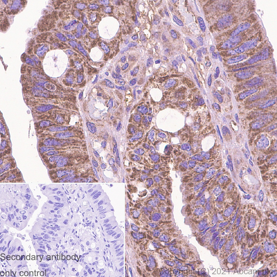
- 5
- 9

Primary Antibodies
AB81298
Anti-FAK (phospho Y397) antibody [EP2160Y]
20ul selling size|RabMAb|Recombinant

- 4
- 2
Reactivity 정보
제품 세부 정보
ab216652 is the carrier-free version of ab32084.
Patented technology
Our RabMAb® technology is a patented hybridoma-based technology for making rabbit monoclonal antibodies. For details on our patents, please refer to RabMAb® patents.
What are the advantages of a recombinant monoclonal antibody?
This product is a recombinant monoclonal antibody, which offers several advantages including:
- - High batch-to-batch consistency and reproducibility
- - Improved sensitivity and specificity
- - Long-term security of supply
- - Animal-free batch production
For more information, read more on recombinant antibodies.
Conjugation ready
Our carrier-free antibodies are typically supplied in a PBS-only formulation, purified and free of BSA, sodium azide and glycerol. This conjugation-ready format is designed for use with fluorochromes, metal isotopes, oligonucleotides, and enzymes, which makes them ideal for antibody labelling, functional and cell-based assays, flow-based assays (e.g. mass cytometry) and Multiplex Imaging applications.
Use our conjugation kits for antibody conjugates that are ready-to-use in as little as 20 minutes with 1 minute hands-on-time and 100% antibody recovery: available for fluorescent dyes, HRP, biotin and gold.
Compatibility
This product is compatible with the Maxpar® Antibody Labeling Kit from Fluidigm, without the need for antibody preparation. Maxpar® is a trademark of Fluidigm Canada Inc.
특성 및 보관 정보
제형
Purification 테크닉
보관 버퍼
배송 시 보관 조건
적절한 단기 보관 조건
적절한 장기 보관 조건
보관 정보
추가 정보
This supplementary information is collated from multiple sources and compiled automatically.
Biological function summary
Paxillin serves key functions in cellular processes such as migration proliferation and survival. It interacts with different proteins including vinculin and talin forming part of a complex at the focal adhesion sites. The phosphorylation state of Paxillin can modulate its interactions and in this way influence the assembly of signaling complexes that control dynamic cellular processes.
Pathways
Paxillin participates in the integrin signaling and MAPK pathways. It operates by linking integrin receptors to the actin cytoskeleton facilitating signal transduction. Paxillin phosphorylation is an important regulatory mechanism within these pathways. In particular its interaction with focal adhesion kinase (FAK) and Src family kinases signifies its role in transmitting signals from the extracellular matrix to the cellular interior which impacts cell behavior and response.
제품 프로토콜
- Visit the General protocols
- Visit the Troubleshooting
타겟 정보
대체 명칭 보기
제품이 사용된 논문 (14)
Recent publications for all applications. Explore the 전체 목록and refine your search
Cell reports 33:108253 PubMed33053339
2020
Applications
Unspecified application
Species
Unspecified reactive species
The Journal of biological chemistry 291:19425-36 PubMed27466370
2016
Applications
Unspecified application
Species
Unspecified reactive species
International journal of cancer 138:1207-19 PubMed26414794
2015
Applications
ICC/IF
Species
Human
PloS one 9:e102424 PubMed25014042
2014
Applications
ICC/IF
Species
Mouse
Journal of translational medicine 11:277 PubMed24180516
2013
Applications
IHC-P
Species
Human
The Journal of biological chemistry 288:35126-37 PubMed24165131
2013
Applications
ICC/IF
Species
Human
Journal of cell science 126:3746-55 PubMed23750011
2013
Applications
Unspecified application
Species
Human
American journal of respiratory cell and molecular biology 49:731-40 PubMed23590307
2013
Applications
Unspecified application
Species
Human papillomavirus
European journal of cell biology 91:171-9 PubMed22284833
2012
Applications
ICC/IF
Species
Unspecified reactive species
The Journal of cell biology 196:223-32 PubMed22270917
2012
Applications
ICC/IF, WB, WB, ICC/IF
Species
Mouse, Mouse, Human, Human
Product promise
Please note: All products are 'FOR RESEARCH USE ONLY. NOT FOR USE IN DIAGNOSTIC OR THERAPEUTIC PROCEDURES'.
For licensing inquiries, please contact partnerships@abcam.com