Anti-Smad1 antibody [EP565Y] - BSA and Azide free
- KO Validated
- RabMAb
- Recombinant
- 각 태그의 뜻
Be the first to review this product! 리뷰 제출
|
(1 출판물)
- ICC/IF
Unknown
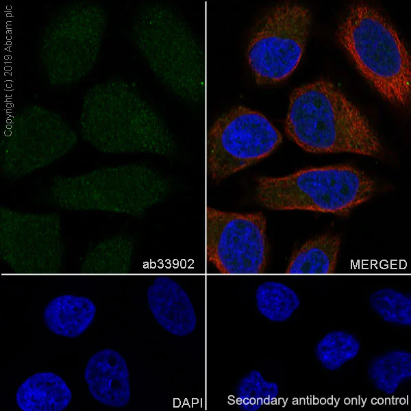
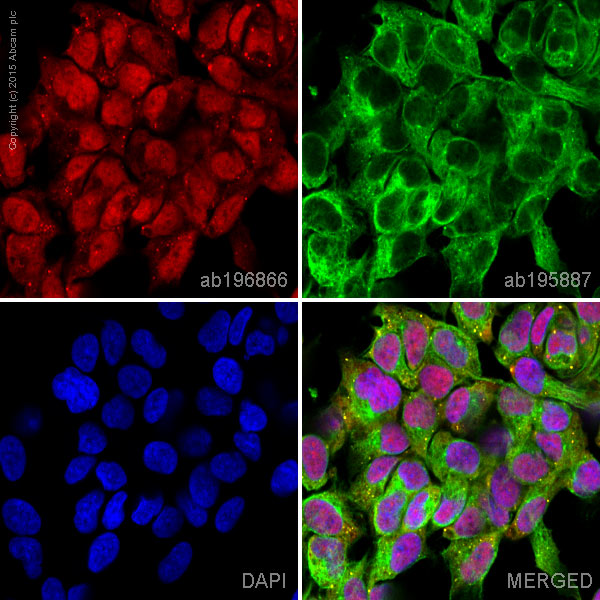
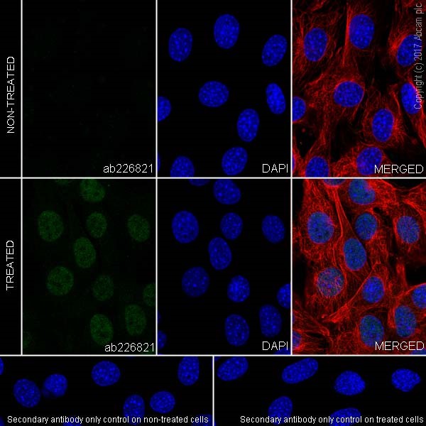
Immunocytochemistry/ Immunofluorescence - Anti-Smad1 antibody [EP565Y] - BSA and Azide free (AB172642)
Immunocytochemistry/ Immunofluorescence analysis of HeLa (Human cervix adenocarcinoma epithelial cell) cells labeling Smad1 with Purified ab33902 at 1/100 dilution (10 µg/mL). Cells were fixed in 4% Paraformaldehyde and permeabilized with 0.1% tritonX-100. Cells were counterstained with ab195889 Anti-alpha Tubulin antibody [DM1A] - Microtubule Marker (Alexa Fluor® 594) 1/200 (2.5 µg/mL). Goat anti rabbit IgG (Alexa Fluor® 488, ab150077) was used as the secondary antibody at 1/1000 (2 µg/mL) dilution. DAPI (blue) was used as nuclear counterstain. PBS instead of the primary antibody was used as the secondary antibody only control.
This data was developed using the same antibody clone in a different buffer formulation containing PBS, BSA, glycerol, and sodium azide (ab33902).
- Flow Cyt (Intra)
Unknown
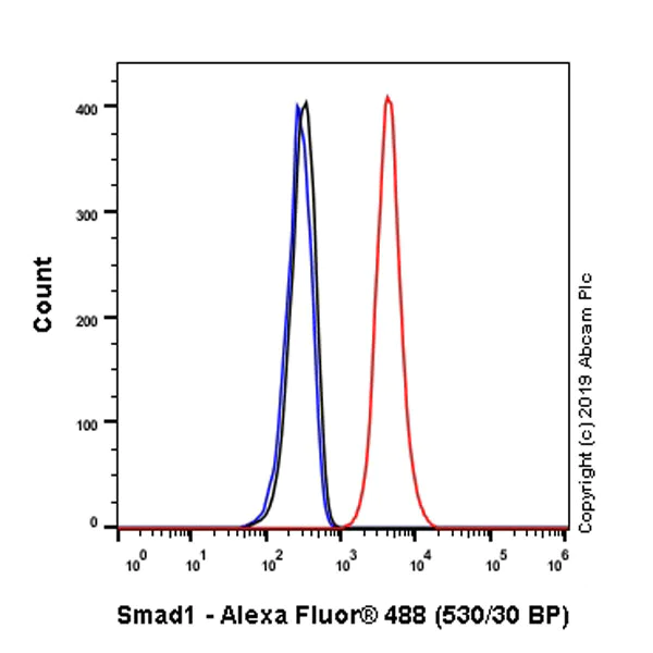
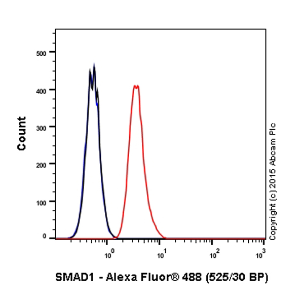
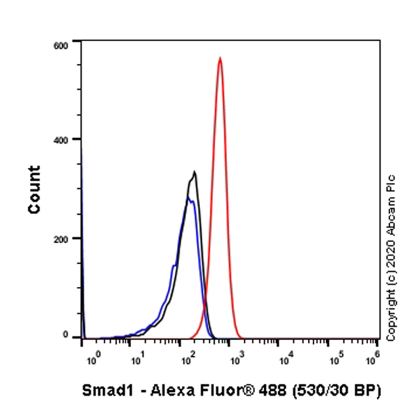
Flow Cytometry (Intracellular) - Anti-Smad1 antibody [EP565Y] - BSA and Azide free (AB172642)
Intracellular Flow Cytometry analysis of HeLa (Human cervix adenocarcinoma epithelial cell) cells labeling Smad1 with Purified ab33902 at 1/100 dilution (10 μg/mL) (Red). Cells were fixed with 4% Paraformaldehyde and permeabilised with 90% Methanol. A Goat anti rabbit IgG (Alexa Fluor® 488, ab150077) secondary antibody was used at 1/2000. Isotype control - Rabbit monoclonal IgG (Black). Unlabeled control - Cell without incubation with primary antibody and secondary antibody (Blue). This data was developed using the same antibody clone in a different buffer formulation containing PBS, BSA, glycerol, and sodium azide (ab33902).
- WB
Unknown
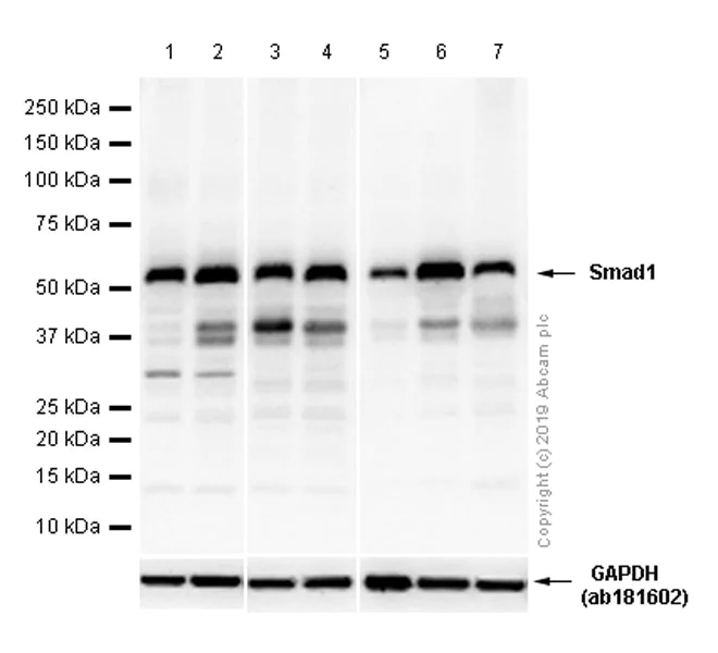
Western blot - Anti-Smad1 antibody [EP565Y] - BSA and Azide free (AB172642)
This data was developed using the same antibody clone in a different buffer formulation containing PBS, BSA, glycerol, and sodium azide (ab33902).
All lanes:
Western blot - Anti-Smad1 antibody [EP565Y] (<a href='/ko/products/primary-antibodies/smad1-antibody-ep565y-ab33902'>ab33902</a>) at 1/1000 dilution
Lane 1:
HEK-293 (Human embryonic kidney epithelial cell) whole cell lysates at 20 µg
Lane 2:
A-673 (Human muscle Ewing's Sarcoma) whole cell lysates at 20 µg
Lane 3:
NIH/3T3 (Mouse embryonic fibroblast) whole cell lysates at 20 µg
Lane 4:
C2C12 (Mouse myoblasts myoblast) whole cell lysates at 20 µg
Lane 5:
Mouse brain lysates at 20 µg
Lane 6:
Mouse skeletal muscle lysates at 20 µg
Lane 7:
Mouse stomach lysates at 20 µg
Secondary
All lanes:
Western blot - Goat Anti-Rabbit IgG H&L (HRP) (<a href='/ko/products/secondary-antibodies/goat-rabbit-igg-h-l-hrp-ab97051'>ab97051</a>) at 1/20000 dilution
Predicted band size: 52 kDa
Observed band size: 58 kDa
false
- WB
Lab
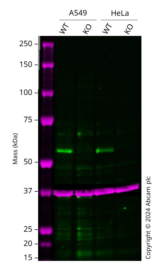
Western blot - Anti-Smad1 antibody [EP565Y] - BSA and Azide free (AB172642)
Western blot : Anti-SMAD1 antibody [EP565Y] (ab33902) staining at 1/1000 dilution, shown in green; Mouse anti-GAPDH antibody [6C5] (ab8245) loading control staining at 1/20000 dilution, shown in magenta. In Western blot, ab33902 was shown to bind specifically to SMAD1. A band was observed at 55-65 kDa in wild-type A549 cell lysates with no signal observed at this size in SMAD1 knockout cell line. To generate this image, wild-type and SMAD1 knockout A549 cell lysates were analysed. First, samples were run on an SDS-PAGE gel then transferred onto a nitrocellulose membrane. Membranes were blocked in 5 % milk in TBS-0.1 % Tween® 20 (TBS-T) before incubation with primary antibodies overnight at 4 °C. Blots were washed four times in TBS-T, incubated with secondary antibodies for 1 h at room temperature, washed again four times then imaged. Secondary antibodies used were Goat anti-Rabbit IgG H&L 800CW and Goat anti-Mouse IgG H&L 680RD at 1/20000 dilution.
All lanes:
Western blot - Anti-Smad1 antibody [EP565Y] (<a href='/ko/products/primary-antibodies/smad1-antibody-ep565y-ab33902'>ab33902</a>) at 1/1000 dilution
Lane 1:
Wild-type A549 cell lysate at 20 µg
Lane 2:
SMAD1 knockout A549 cell lysate at 20 µg
Lane 3:
Wild-type HeLa ab255929 cell lysate at 20 µg
Lane 4:
SMAD1 knockout HeLa <a href='/ko/products/cell-lines/human-smad1-knockout-hela-cell-line-ab265400'>ab265400</a> cell lysate at 20 µg
Secondary
All lanes:
Goat anti-Rabbit IgG H&L 800CW and Goat anti-Mouse IgG H&L 680RD at 1/20000 dilution
false
관련 conjugated 항체와 다양한 조성의 항체 (1)
-
Anti-Smad1 antibody [EP565Y]
대체품
Primary Antibodies
AB214423
Anti-Smad1 (phospho S463 + S465) antibody [EPR20662-29]
20ul selling size|RabMAb|Recombinant|Trial Size
- 0
- 0
Primary Antibodies
AB226821
Anti-Smad1 (phospho S463 + S465) antibody [EPR20662-20]
20ul selling size|RabMAb|Recombinant
- 0
- 0
Primary Antibodies
AB126761
20ul selling size|KO Validated|RabMAb|Recombinant
- 0
- 0
Primary Antibodies
AB228457
Anti-Smad1 (phospho S463 + S465) antibody [EPR20662-20] - BSA and Azide free
RabMAb|Recombinant
- 0
- 0
보완 제품
Primary Antibodies
AB40771
20ul selling size|KO Validated|RabMAb|Recombinant
- 5
- 4
Primary Antibodies
AB92698
Anti-SMAD1 + SMAD5 + SMAD9 (phospho S463 + S465 + S467) antibody [MMC-1-104-3]
20ul selling size|RabMAb|Recombinant
- 4
- 2
Primary Antibodies
AB16667
BOND RX™ Validated|KO Validated|RabMAb|Advanced Validation|Recombinant|20ul selling size

- 5
- 133
Primary Antibodies
AB226821
Anti-Smad1 (phospho S463 + S465) antibody [EPR20662-20]
20ul selling size|RabMAb|Recombinant
- 0
- 0
Reactivity 정보
제품 세부 정보
ab172642 is the carrier-free version of ab33902.
Patented technology
Our RabMAb® technology is a patented hybridoma-based technology for making rabbit monoclonal antibodies. For details on our patents, please refer to RabMAb® patents.
What are the advantages of a recombinant monoclonal antibody?
This product is a recombinant monoclonal antibody, which offers several advantages including:
- - High batch-to-batch consistency and reproducibility
- - Improved sensitivity and specificity
- - Long-term security of supply
- - Animal-free batch production
For more information, read more on recombinant antibodies.
Conjugation ready
Our carrier-free antibodies are typically supplied in a PBS-only formulation, purified and free of BSA, sodium azide and glycerol. This conjugation-ready format is designed for use with fluorochromes, metal isotopes, oligonucleotides, and enzymes, which makes them ideal for antibody labelling, functional and cell-based assays, flow-based assays (e.g. mass cytometry) and Multiplex Imaging applications.
Use our conjugation kits for antibody conjugates that are ready-to-use in as little as 20 minutes with 1 minute hands-on-time and 100% antibody recovery: available for fluorescent dyes, HRP, biotin and gold.
Compatibility
This product is compatible with the Maxpar® Antibody Labeling Kit from Fluidigm, without the need for antibody preparation. Maxpar® is a trademark of Fluidigm Canada Inc.
특성 및 보관 정보
제형
Purification 테크닉
보관 버퍼
배송 시 보관 조건
적절한 단기 보관 조건
적절한 장기 보관 조건
보관 정보
추가 정보
This supplementary information is collated from multiple sources and compiled automatically.
Biological function summary
Smad1 participates in the transmission of BMP signals from the cell surface to the nucleus ensuring transcriptional regulation of target genes. In the presence of BMPs Smad1 forms complexes with Smad4 upon activation. These Smad complexes then translocate to the nucleus where they regulate gene expression. Smad1 influences cellular responses such as proliferation differentiation and apoptosis. In particular it plays a significant role in bone development and osteogenesis.
Pathways
Smad1 plays an important role within the BMP signaling pathway which is important for early development and tissue homeostasis. Within this pathway BMPs trigger the phosphorylation of Smad1 which then associates with Smad4 to propagate downstream signaling. Another associated pathway includes the TGF-beta signaling pathway where Smads like Smad2 and Smad3 show functional similarities and differences with Smad1. The interactions of Smad1 with related proteins like Smad4 highlight its significant contribution to cellular processes regulated by these pathways.
제품 프로토콜
- Visit the General protocols
- Visit the Troubleshooting
타겟 정보
대체 명칭 보기
제품이 사용된 논문 (1)
Recent publications for all applications. Explore the 전체 목록and refine your search
The Journal of biological chemistry 300:107988 PubMed39542244
2024
Applications
Unspecified application
Species
Unspecified reactive species
Product promise
Please note: All products are 'FOR RESEARCH USE ONLY. NOT FOR USE IN DIAGNOSTIC OR THERAPEUTIC PROCEDURES'.
For licensing inquiries, please contact partnerships@abcam.com