Anti-SMAD3 (pS423/425)+SMAD5&SMAD1 (pS463/465)+SMAD2 (pS465/467) antibody [EP823Y] - BSA&Azide free
- Recombinant
- Advanced Validation
- RabMAb
- 각 태그의 뜻
3
(1 리뷰)
|
(2 제품이 사용된 논문 )

- IHC-P
Unknown
Immunohistochemistry (Formalin/PFA-fixed paraffin-embedded sections) - Anti-SMAD3 (pS423/425) + SMAD5 (pS463/465 ) + SMAD1 (pS463/465) + SMAD2 (pS465/467) antibody [EP823Y] - BSA and Azide free (AB172202)
Immunohistochemical analysis of Smad3 in paraffin embedded human liver carcinoma tissue using ab52903 at 1/100 dilution.This data was developed using the same antibody clone in a different buffer formulation containing PBS, BSA, glycerol, and sodium azide (ab52903).

- ICC/IF
Unknown
Immunocytochemistry/ Immunofluorescence - Anti-SMAD3 (pS423/425)+SMAD5&SMAD1 (pS463/465)+SMAD2 (pS465/467) antibody [EP823Y] - BSA&Azide free (AB172202)
Immunocytochemistry/Immunofluorescence analysis of A549 +/- TGFβ (5ng/ml 24h) and A549 + TGFβ (5ng/ml 24h) + Lamda phosphatase (LP) cells. Smad3 (phospho S423 + S425) was labelled with purified ab52903 at a dilution of 1/100 dilution while Smad3 was labelled with ab207447 at a dilution of 1/500 dilution. Cells were fixed with 4% paraformaldehyde and permeabilized with 0.1% triton X-100. ab150077 (goat anti-rabbit IgG Alexa Fluor® 488) (1/1000 dilution) was used as the secondary antibody. The cells were co-stained with ab195889 (Anti-alpha Tubulin antibody [DM1A] - Microtubule Marker (Alexa Fluor® 594)) at 1/200 dilution. Nuclei counterstained with DAPI (blue). Control : PBS instead of the primary antibody.
This ICC/IF data was generated using the same anti-phospho Smad3 S423/425 antibody clone, EP823Y, in a different buffer formulation (ab52903).

- ICC/IF
AbReview40862****
Immunocytochemistry/ Immunofluorescence - Anti-SMAD3 (pS423/425)+SMAD5&SMAD1 (pS463/465)+SMAD2 (pS465/467) antibody [EP823Y] - BSA&Azide free (AB172202)
ab52903 staining Smad3 in mouse primary embryonic epicardial cells by ICC/IF (Immunocytochemistry/immunofluorescence). Cells were fixed with 4% formaldehyde permeabilized with 0.5% Triton X-100 and blocked with PBS + 1% BSA + 10% goat serum + 0.1% Triton X-100 for 1 hour at 20°C. Samples were incubated with primary antibody (1/100 dilution in PBS + 1% BSA + 10% goat serum + 0.1% Triton X-100) for 16 hours at 4°C. An Alexa Fluor®488-conjugated goat anti-rabbit IgG polyclonal (1/200 dilution) was used as the secondary antibody.
This data was developed using the same antibody clone in a different buffer formulation containing PBS, BSA, glycerol, and sodium azide (ab52903).
This image is courtesy of an anonymous Abreview.

- IHC-P
Unknown
Immunohistochemistry (Formalin/PFA-fixed paraffin-embedded sections) - Anti-SMAD3 (pS423/425) + SMAD5 (pS463/465 ) + SMAD1 (pS463/465) + SMAD2 (pS465/467) antibody [EP823Y] - BSA and Azide free (AB172202)
This IHC data was generated using the same anti-phospho Smad3 S423/425 antibody clone, EP823Y, in a different buffer formulation (cat# ab52903).
ab52903 staining Smad3 in Human stomach tissue sections by Immunohistochemistry (Formalin/PFA fixed paraffin embedded sections). Tissue was fixed with paraffin and antigen retrieval was by heat mediation using ab93684 (Tris/EDTA buffer, Ph9.0). Samples were incubated with primary antibody at a 1/200 dilution. A ready to use rabbit specific IHC polymer detection kit HRP/DAP (ab209101). Hematoxylin was used as a counterstain. Nuclear and weakly cytoplasmic staining on human stomach without alkaline phosphatase treatment (image A). No signal can be detected when tissues were treated with alkaline phosphatase (image B).

- ICC/IF
AbReview16498****
Immunocytochemistry/ Immunofluorescence - Anti-SMAD3 (pS423/425)+SMAD5&SMAD1 (pS463/465)+SMAD2 (pS465/467) antibody [EP823Y] - BSA&Azide free (AB172202)
ab52903 staining Smad3 (phospho S423 + S425) in human TII Pneumocyte A549 cells by Immunocytochemistry/ Immunofluorescence. Cells were fixed with paraformaldehyde and permeabilized with 0.1% Triton x100 before blocking with 3% BSA for 1 hour at RT. Samples were incubated with primary antibody (1/200 : in 3% BSA in 1x PBST) for 24 hours at 4°C. A TRITC-conjugated goat polyclonal to rabbit IgG was used as secondary antibody at 1/200 dilution.
This data was developed using the same antibody clone in a different buffer formulation containing PBS, BSA, glycerol, and sodium azide (ab52903).
This image is courtesy of an Abreview submitted by Aaron Gardner.

- IHC-P
Unknown
Immunohistochemistry (Formalin/PFA-fixed paraffin-embedded sections) - Anti-SMAD3 (pS423/425) + SMAD5 (pS463/465 ) + SMAD1 (pS463/465) + SMAD2 (pS465/467) antibody [EP823Y] - BSA and Azide free (AB172202)
Purified ab52903 staining Smad3 in Mouse kidney tissue sections by Immunohistochemistry (Formalin/PFA fixed paraffin embedded sections). Tissue was fixed with paraffin and antigen retrieval was by heat mediation using ab93684 (Tris/EDTA buffer, pH 9.0). Samples were incubated with primary antibody at a 1/200 dilution. A ready to use rabbit specific IHC polymer detection kit HRP/DAP (ab209101). Hematoxylin was used as a counterstain. Nuclear and weakly cytoplasmic staining on mouse kidney without alkaline phosphatase treatment (image A). No signal can be detected when tissues were treated with alkaline phosphatase (image B).
This data was developed using the same antibody clone in a different buffer formulation containing PBS, BSA, glycerol, and sodium azide (ab52903).

- ICC/IF
PubMed
Immunocytochemistry/ Immunofluorescence - Anti-SMAD3 (pS423/425)+SMAD5&SMAD1 (pS463/465)+SMAD2 (pS465/467) antibody [EP823Y] - BSA&Azide free (AB172202)
TGF-β1 signaling is impaired in NDRG1-silenced MEFs. PML+/+ mouse embryonic fibroblasts (MEFs) were transfected with either CTL-siRNAs (A & B) or NDRG1-siRNAs (C & D) and induced with100 ng/ml TGF-β1. Immunofluorescent staining revealed intense nuclear staining for phosphorylated SMAD3 (SMAD3-P ab52903) in CTL-siRNA treated MEFs (B) while only weak nuclear staining for MEFs treated with NDRG1-siRNA (D).
This data was developed using the same antibody clone in a different buffer formulation containing PBS, BSA, glycerol, and sodium azide (ab52903).
Image from Tang MK et al. PLoS One. 2013;8(3):e59477. Fig 12.; doi: 10.1371/journal.pone.0059477.

- Dot
Unknown
Dot Blot - Anti-SMAD3 (pS423/425)+SMAD5&SMAD1 (pS463/465)+SMAD2 (pS465/467) antibody [EP823Y] - BSA&Azide free (AB172202)
Dot blot analysis of Smad 3 (phospho S423 + S425) phospho peptide (Lane 1), Smad 3 (phospho S423) phospho peptide (Lane 2), Smad 3 (phospho S425) phospho peptide (Lane 3) and Smad 3 non-phospho peptide (Lane 4) labelling Smad 3 (phospho S423 + S425) with ab52903 at a dilution of 1/1000. A Goat Anti-Rabbit IgG, (H+L), Peroxidase conjugated (ab97051) was used as the secondary antibody at a dilution of 1/20,000. Blocking and dilution buffer : 5% NFDM /TBST.
This data was developed using the same antibody clone in a different buffer formulation containing PBS, BSA, glycerol, and sodium azide (ab52903).

- ChIC/CUT&RUN-seq
Lab
ChIC/CUT&RUN sequencing - Anti-SMAD3 (pS423/425)+SMAD5&SMAD1 (pS463/465)+SMAD2 (pS465/467) antibody [EP823Y] - BSA&Azide free (AB172202)
This data was developed using the same antibody clone in a different buffer formulation (ab52903). ChIC/CUT&RUN was performed using a pAG-MNase at a final concentration of 700 ng/mL, 2.5 x 10^5 A549 (Human lung carcinoma cell line) cells treated with hTGF-β1 (7 ng/mL 1 h) and 5 µg of ab52903 [EP823Y]. The resulting DNA was sequenced on the Illumina NovaSeq 6000 to a depth of 10 million reads. The negative IgG control ab172730 is also shown. The University of Geneva owns patents relevant to ChIC (Chromatin Immuno-Cleavage) methods.

- IHC-P
PubMed
Immunohistochemistry (Formalin/PFA-fixed paraffin-embedded sections) - Anti-SMAD3 (pS423/425) + SMAD5 (pS463/465 ) + SMAD1 (pS463/465) + SMAD2 (pS465/467) antibody [EP823Y] - BSA and Azide free (AB172202)
Representative IHC photomicrographs from an Environmental enteropathy (EE) duodenal biopsy showing p-SMAD3 staining (ab52903) in only the epithelium (arrows).
This data was developed using the same antibody clone in a different buffer formulation containing PBS, BSA, glycerol, and sodium azide (ab52903).
Image from Syed S et al. PLoS Negl Trop Dis. 2018;12(2):e0006224. Fig 4.; doi: 10.1371/journal.pntd.0006224.

- Dot
Lab
Dot Blot - Anti-SMAD3 (pS423/425)+SMAD5&SMAD1 (pS463/465)+SMAD2 (pS465/467) antibody [EP823Y] - BSA&Azide free (AB172202)
Blocking and diluting buffer and concentration : 5% NFDM /TBST
All lanes:
Dot Blot - Anti-SMAD3 (pS423/425) + SMAD5 (pS463/465) + SMAD1 (pS463/465) + SMAD2 (pS465/467) antibody [EP823Y] (<a href='/ko/products/primary-antibodies/smad3-ps423-425-smad5-ps463-465-smad1-ps463-465-smad2-ps465-467-antibody-ep823y-ab52903'>ab52903</a>) at 1/1000 dilution
Lane 1:
Smad5 non-phospho peptide
Lane 2:
Smad5 (s463+s465) phospho peptide
Lane 3:
Smad2 non-phospho peptide
Lane 4:
Smad2 (s465+s467) phospho peptide
Lane 5:
Smad1 non-phospho peptide
Lane 6:
Smad1 (s463+s465) phospho peptide
Lane 7:
Smad3 non-phospho peptide
Lane 8:
Smad3 (s423+s435) phospho peptide
Secondary
All lanes:
Dot Blot - Goat Anti-Rabbit IgG H&L (HRP) (<a href='/ko/products/secondary-antibodies/goat-rabbit-igg-h-l-hrp-ab97051'>ab97051</a>) at 1/20000 dilution
false
Exposure time: 59s
관련 conjugated 항체와 다양한 조성의 항체 (7)
-
Anti-SMAD3 (pS423/425) + SMAD5 (pS463/465) + SMAD1 (pS463/465) + SMAD2 (pS465/467) antibody [EP823Y]
-
519 Alexa Fluor® 488
AF488 Anti-SMAD3 (pS423/425) + SMAD5 & SMAD1 (pS463/465) + SMAD2 (pS465/467) antibody [EP823Y]
-
565 Alexa Fluor® 555
AF555 Anti-SMAD3 (pS423/425) + SMAD5 & SMAD1 (pS463/465) + SMAD2 (pS465/467) antibody [EP823Y]
-
603 Alexa Fluor® 568
AF568 Anti-SMAD3 (pS423/425) + SMAD5 & SMAD1 (pS463/465) + SMAD2 (pS465/467) antibody [EP823Y]
-
617 Alexa Fluor® 594
AF594 Anti-SMAD3 (pS423/425) + SMAD5 & SMAD1 (pS463/465) + SMAD2 (pS465/467) antibody [EP823Y]
-
665 Alexa Fluor® 647
AF647 Anti-SMAD3 (pS423/425) + SMAD5 & SMAD1 (pS463/465) + SMAD2 (pS465/467) antibody [EP823Y]
-
775 Alexa Fluor® 750
A750 Anti-SMAD3 (pS423/425)+SMAD5 (pS463/465)+SMAD1 (pS463/465)+SMAD2 (pS465/467) antibody [EP823Y]
대체품


Primary Antibodies
AB40854
20ul selling size|Recombinant|KO Validated|Advanced Validation|RabMAb

- 5
- 10
Primary Antibodies
AB157372
Anti-Smad3 antibody [EP568Y] - BSA and Azide free
Recombinant|KO Validated|Advanced Validation|RabMAb

- 0
- 0


Primary Antibodies
AB208182
Anti-Smad3 antibody [EPR19686] - ChIP Grade
20ul selling size|RabMAb|Recombinant|KO Validated

- 0
- 0
보완 제품
Sample Prep & Detection Kits
AB269901
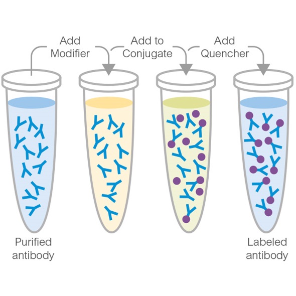
Alkaline Phosphatase Conjugation Kit (Animal Free) - Lightning-Link®
- 0
- 0

Primary Antibodies
AB40854
20ul selling size|Recombinant|KO Validated|Advanced Validation|RabMAb
- 5
- 10
Primary Antibodies
AB215715
Anti-TGF beta 1 antibody [EPR21143]
20ul selling size|BOND RX™ Validated|RabMAb|Recombinant|KO Validated

- 5
- 7
Primary Antibodies
AB280888
Anti-Smad2 (phospho S467) antibody [EPR23681-40]
BOND RX™ Validated|20ul selling size|RabMAb|Recombinant|KO Validated

- 5
- 2
Primary Antibodies
AB124964
Anti-alpha smooth muscle Actin antibody [EPR5368]
BOND RX™ Validated|RabMAb|Recombinant|KO Validated|20ul selling size

- 5
- 19
Reactivity 정보
제품 세부 정보
ab172202 is the carrier-free version of ab52903.
Patented technology
Our RabMAb® technology is a patented hybridoma-based technology for making rabbit monoclonal antibodies. For details on our patents, please refer to RabMAb® patents.
What are the advantages of a recombinant monoclonal antibody?
This product is a recombinant monoclonal antibody, which offers several advantages including:
- - High batch-to-batch consistency and reproducibility
- - Improved sensitivity and specificity
- - Long-term security of supply
- - Animal-free batch production
For more information, read more on recombinant antibodies.
Conjugation ready
Our carrier-free antibodies are typically supplied in a PBS-only formulation, purified and free of BSA, sodium azide and glycerol. This conjugation-ready format is designed for use with fluorochromes, metal isotopes, oligonucleotides, and enzymes, which makes them ideal for antibody labelling, functional and cell-based assays, flow-based assays (e.g. mass cytometry) and Multiplex Imaging applications.
Use our conjugation kits for antibody conjugates that are ready-to-use in as little as 20 minutes with 1 minute hands-on-time and 100% antibody recovery: available for fluorescent dyes, HRP, biotin and gold.
Compatibility
This product is compatible with the Maxpar® Antibody Labeling Kit from Fluidigm, without the need for antibody preparation. Maxpar® is a trademark of Fluidigm Canada Inc.
특성 및 보관 정보
제형
Purification 테크닉
보관 버퍼
배송 시 보관 조건
적절한 단기 보관 조건
적절한 장기 보관 조건
보관 정보
추가 정보
This supplementary information is collated from multiple sources and compiled automatically.
Biological function summary
Smad3 acts as a mediator of signal transduction for the TGF-beta (transforming growth factor-beta) superfamily forming a complex with phosphorylated Smad2. This enables it to regulate transcriptional activity influencing cell proliferation differentiation and apoptosis. Smad3 also participates in various cellular processes by interacting with other co-factors and regulatory proteins that aid in fine-tuning its function.
Pathways
Smad3 plays an important role in the TGF-beta signaling pathway where it works closely with Smad4 to propagate the signal. Upon phosphorylation it forms a complex with co-Smad (Smad4) and moves into the nucleus to influence gene expression. Smad3 is also involved in pathways related to oncogenesis and tissue fibrosis indicating its significant role in cellular regulation and response mechanisms.
제품 프로토콜
- Visit the General protocols
- Visit the Troubleshooting
타겟 정보
Additional targets
대체 명칭 보기
제품이 사용된 논문 (2)
Recent publications for all applications. Explore the 전체 목록and refine your search
Journal of Cancer 14:114-128 PubMed36605486
2023
Applications
Unspecified application
Species
Unspecified reactive species
The European journal of neuroscience 54:5586-5600 PubMed34258805
2021
Applications
Unspecified application
Species
Unspecified reactive species
Product promise
Please note: All products are 'FOR RESEARCH USE ONLY. NOT FOR USE IN DIAGNOSTIC OR THERAPEUTIC PROCEDURES'.
For licensing inquiries, please contact partnerships@abcam.com