Anti-mH2A1 antibody (ab37264)
")
Key features and details
- Rabbit polyclonal to mH2A1
- Suitable for: ChIP, WB, IHC-P, ICC/IF
- Knockout validated
- Reacts with: Mouse, Human
- Isotype: IgG
Get better batch-to-batch reproducibility with a recombinant antibody

- Research with confidence – consistent and reproducible results with every batch
- Long-term and scalable supply – powered by recombinant technology for fast production
- Success from the first experiment – confirmed specificity through extensive validation
- Ethical standards compliant – production is animal-free
Overview
-
Product name
Anti-mH2A1 antibody
See all mH2A1 primary antibodies -
Description
Rabbit polyclonal to mH2A1 -
Host species
Rabbit -
Specificity
ab37264 recognises the three known isoforms of mH2A1 including mH2A1.2 (longest isoform) and the mH2A1.1 (shortest isoform).
We have had varying reports about the efficiency with which this antibody recognises mH2A1 in mouse cells and tissues. Please contact our Scientific Support team if you have any queries about this. -
Tested applications
Suitable for: ChIP, WB, IHC-P, ICC/IFmore details -
Species reactivity
Reacts with: Mouse, Human
Predicted to work with: Rat, Chicken, Cow, Xenopus laevis
-
Immunogen
Synthetic peptide corresponding to Human mH2A1 aa 150-250 conjugated to keyhole limpet haemocyanin.
(Peptide available asab37263) -
Positive control
- ICC/IF: HeLa cells.
-
General notes
The Life Science industry has been in the grips of a reproducibility crisis for a number of years. Abcam is leading the way in addressing this with our range of recombinant monoclonal antibodies and knockout edited cell lines for gold-standard validation. Please check that this product meets your needs before purchasing.
If you have any questions, special requirements or concerns, please send us an inquiry and/or contact our Support team ahead of purchase. Recommended alternatives for this product can be found below, along with publications, customer reviews and Q&As
Properties
-
Form
Liquid -
Storage instructions
Shipped at 4°C. Store at +4°C short term (1-2 weeks). Upon delivery aliquot. Store at -20°C or -80°C. Avoid freeze / thaw cycle. -
Storage buffer
pH: 7.40
Preservative: 0.02% Sodium azide
Constituent: PBS
Batches of this product that have a concentration < 1mg/ml may have BSA added as a stabilising agent. If you would like information about the formulation of a specific lot, please contact our scientific support team who will be happy to help. -
 Concentration information loading...
 -
Purity
Immunogen affinity purified -
Clonality
Polyclonal -
Isotype
IgG -
Research areas
Associated products
-
ChIP Related Products
-
Compatible Secondaries
-
Isotype control
-
Recombinant Protein
Applications
The Abpromise guarantee
Our Abpromise guarantee covers the use of ab37264 in the following tested applications.
The application notes include recommended starting dilutions; optimal dilutions/concentrations should be determined by the end user.
| Application | Abreviews | Notes |
|---|---|---|
| ChIP |
Use at an assay dependent concentration. PubMed: 19380460
|
|
| WB | (1) |
Use a concentration of 1 µg/ml. Detects a band of approximately 40 kDa (predicted molecular weight: 40 kDa).
|
| IHC-P | (1) |
Use a concentration of 5 µg/ml. Perform heat mediated antigen retrieval before commencing with IHC staining protocol.
|
| ICC/IF | (1) |
Use a concentration of 1 - 5 µg/ml.
|
| Notes |
|---|
|
ChIP
Use at an assay dependent concentration. PubMed: 19380460 |
|
WB
Use a concentration of 1 µg/ml. Detects a band of approximately 40 kDa (predicted molecular weight: 40 kDa). |
|
IHC-P
Use a concentration of 5 µg/ml. Perform heat mediated antigen retrieval before commencing with IHC staining protocol. |
|
ICC/IF
Use a concentration of 1 - 5 µg/ml. |
Target
-
Function
Variant histone H2A which replaces conventional H2A in a subset of nucleosomes where it represses transcription. Nucleosomes wrap and compact DNA into chromatin, limiting DNA accessibility to the cellular machineries which require DNA as a template. Histones thereby play a central role in transcription regulation, DNA repair, DNA replication and chromosomal stability. DNA accessibility is regulated via a complex set of post-translational modifications of histones, also called histone code, and nucleosome remodeling. Involved in stable X chromosome inactivation. Inhibits the binding of transcription factors and interferes with the activity of remodeling SWI/SNF complexes. Inhibits histone acetylation by EP300 and recruits class I HDACs, which induces an hypoacetylated state of chromatin. In addition, isoform 1, but not isoform 2, binds ADP-ribose and O-acetyl-ADP-ribose, and may be involved in ADP-ribose-mediated chromatin modulation. -
Tissue specificity
Ubiquitous. -
Sequence similarities
Contains 1 histone H2A domain.
Contains 1 Macro domain. -
Post-translational
modificationsMonoubiquitinated at either Lys-116 or Lys-117. May also be polyubiquitinated. Ubiquitination is mediated by the CUL3/SPOP E3 complex and does not promote proteasomal degradation. Instead, it is required for enrichment in inactive X chromosome chromatin. -
Cellular localization
Nucleus. Chromosome. Enriched in inactive X chromosome chromatin and in senescence-associated heterochromatin. - Information by UniProt
-
Database links
- Entrez Gene: 9555 Human
- Entrez Gene: 26914 Mouse
- Entrez Gene: 29384 Rat
- Omim: 610054 Human
- SwissProt: O75367 Human
- SwissProt: Q9QZQ8 Mouse
- SwissProt: Q02874 Rat
- Unigene: 420272 Human
see all -
Alternative names
- Core histone macro h2a.1 antibody
- Core histone macro-H2A.1 antibody
- H2A histone family member Y antibody
see all
Images
-
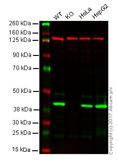
Western blot - Anti-mH2A1 antibody (ab37264)
Lane 1: Wild-type HAP1 whole cell lysate (20 µg)
Lane 2: MH2A1 knockout HAP1 whole cell lysate (20 µg)
Lane 3: HeLa whole cell lysate (20 µg)
Lane 4: HepG2 whole cell lysate (20 µg)Lanes 1 - 4: Merged signal (red and green). Green - ab37264 observed at 40 kDa. Red - loading control, ab18058, observed at 130 kDa.
ab37264 was shown to specifically recognize MH2A1 in wild-type HAP1 cells along with additional cross reactive bands. No band was observed when MH2A1 knockout cells were examined. Wild-type and MH2A1 knockout samples were subjected to SDS-PAGE. Ab37264 and ab18058 (Mouse anti Vinculin loading control) were incubated overnight at 4°C at 1 ug/ml and 1/10,000 dilution respectively. Blots were developed with Goat anti-Rabbit IgG H&L (IRDye® 800CW) preabsorbed (ab216773) and Goat anti-Mouse IgG H&L (IRDye® 680RD) preabsorbed (ab216776) secondary antibodies at 1/10,000 dilution for 1 hour at room temperature before imaging.
-
 Immunocytochemistry/ Immunofluorescence - Anti-mH2A1 antibody (ab37264)
ab37264 staining mH2A1 in HeLa cells. The cells were fixed with 4% paraformaldehyde (10 min), permeabilized with 0.1% PBS-Triton X-100 for 5 minutes and then blocked with 1% BSA/10% normal goat serum/0.3M glycine in 0.1%PBS-Tween for 1h. The cells were then incubated overnight at 4°C with ab37264 at 1µg/ml and ab7291, Mouse monoclonal [DM1A] to alpha Tubulin - Loading Control. Cells were then incubated with ab150081, Goat polyclonal Secondary Antibody to Rabbit IgG - H&L (Alexa Fluor® 488), pre-adsorbed at 1/1000 dilution (shown in green) and ab150120, Goat polyclonal Secondary Antibody to Mouse IgG - H&L (Alexa Fluor® 594), pre-adsorbed at 1/1000 dilution (shown in pseudocolour red). Nuclear DNA was labelled with DAPI (shown in blue).
Also suitable in cells fixed with 100% methanol (5 min).
Image was acquired with a high-content analyser (Operetta CLS, Perkin Elmer) and a maximum intensity projection of confocal sections is shown.
-
 Immunohistochemistry (Formalin/PFA-fixed paraffin-embedded sections) - Anti-mH2A1 antibody (ab37264)
IHC image of mH2A1 staining in human skin FFPE section, performed on a Leica BondTM system using the standard protocol F. The section was pre-treated using heat mediated antigen retrieval with sodium citrate buffer (pH6, epitope retrieval solution 1) for 20 mins. The section was then incubated with ab37264, 5µg/ml, for 15 mins at room temperature and detected using an HRP conjugated compact polymer system. DAB was used as the chromogen. The section was then counterstained with haematoxylin and mounted with DPX.
-
 Western blot - Anti-mH2A1 antibody (ab37264)All lanes : Anti-mH2A1 antibody (ab37264) at 1 µg
Lane 1 : HeLa (Human epithelial carcinoma cell line) Whole Cell Lysate
Lane 2 : HepG2 (Human hepatocellular liver carcinoma cell line) Whole Cell Lysate
Lane 3 : NIH 3T3 (Mouse embryonic fibroblast cell line) Whole Cell Lysate
Lysates/proteins at 20 µg per lane.
Secondary
All lanes : Goat Anti-Rabbit IgG H&L (HRP) (ab97051) at 1/10000 dilution
Developed using the ECL technique.
Performed under reducing conditions.
Predicted band size: 40 kDa
Observed band size: 40 kDa
Additional bands at: 100 kDa. We are unsure as to the identity of these extra bands.
Exposure time: 16 minutes
Protocols
Datasheets and documents
-
SDS download
-
Datasheet download
References (32)
ab37264 has been referenced in 32 publications.
- Han D et al. A balanced Oct4 interactome is crucial for maintaining pluripotency. Sci Adv 8:eabe4375 (2022). PubMed: 35171666
- Oliviero G et al. Distinct and diverse chromatin proteomes of ageing mouse organs reveal protein signatures that correlate with physiological functions. Elife 11:N/A (2022). PubMed: 35259090
- Xu X et al. The epigenetic regulator LSH maintains fork protection and genomic stability via MacroH2A deposition and RAD51 filament formation. Nat Commun 12:3520 (2021). PubMed: 34112784
- Kumbhar R et al. Poly(ADP-ribose) binding and macroH2A mediate recruitment and functions of KDM5A at DNA lesions. J Cell Biol 220:N/A (2021). PubMed: 34003252
- Ma H et al. MacroH2A1.2 deficiency leads to neural stem cell differentiation defects and autism-like behaviors. EMBO Rep 22:e52150 (2021). PubMed: 34046991