Anti-NSMase2 antibody (ab85017)
")
Key features and details
- Rabbit polyclonal to NSMase2
- Suitable for: ICC/IF, WB
- Reacts with: Rat
- Isotype: IgG
Overview
-
Product name
Anti-NSMase2 antibody
See all NSMase2 primary antibodies -
Description
Rabbit polyclonal to NSMase2 -
Host species
Rabbit -
Tested applications
Suitable for: ICC/IF, WBmore details -
Species reactivity
Reacts with: Rat
Predicted to work with: Mouse, Human
-
Immunogen
Synthetic peptide. This information is proprietary to Abcam and/or its suppliers.
-
Positive control
- WB: Rat brain tissue lysate. IHC-P: Human lung tissue. ICC/IF: Primary hippocampal rat neurons/glia, (obtained from Neuromics, cat. no. PC35101), DIV14.
-
General notes
The Life Science industry has been in the grips of a reproducibility crisis for a number of years. Abcam is leading the way in addressing this with our range of recombinant monoclonal antibodies and knockout edited cell lines for gold-standard validation. Please check that this product meets your needs before purchasing.
If you have any questions, special requirements or concerns, please send us an inquiry and/or contact our Support team ahead of purchase. Recommended alternatives for this product can be found below, along with publications, customer reviews and Q&As
Properties
-
Form
Liquid -
Storage instructions
Shipped at 4°C. Store at +4°C short term (1-2 weeks). Upon delivery aliquot. Store at -20°C or -80°C. Avoid freeze / thaw cycle. -
Storage buffer
pH: 7.40
Preservative: 0.02% Sodium azide
Constituent: PBS
1x PBS -
 Concentration information loading...
 -
Purity
Immunogen affinity purified -
Clonality
Polyclonal -
Isotype
IgG -
Research areas
Associated products
-
Compatible Secondaries
-
Isotype control
-
Recombinant Protein
Applications
The Abpromise guarantee
Our Abpromise guarantee covers the use of ab85017 in the following tested applications.
The application notes include recommended starting dilutions; optimal dilutions/concentrations should be determined by the end user.
| Application | Abreviews | Notes |
|---|---|---|
| ICC/IF | (2) |
Use a concentration of 5 µg/ml.
|
| WB |
Use a concentration of 1 µg/ml. Detects a band of approximately 71 kDa (predicted molecular weight: 71 kDa).
|
| Notes |
|---|
|
ICC/IF
Use a concentration of 5 µg/ml. |
|
WB
Use a concentration of 1 µg/ml. Detects a band of approximately 71 kDa (predicted molecular weight: 71 kDa). |
Target
-
Function
Catalyzes the hydrolysis of sphingomyelin to form ceramide and phosphocholine. Ceramide mediates numerous cellular functions, such as apoptosis and growth arrest, and is capable of regulating these 2 cellular events independently. Also hydrolyzes sphingosylphosphocholine. Regulates the cell cycle by acting as a growth suppressor in confluent cells. Probably acts as a regulator of postnatal development and participates in bone and dentin mineralization. -
Tissue specificity
Predominantly expressed in brain. -
Sequence similarities
Belongs to the neutral sphingomyelinase family. -
Developmental stage
Up-regulated during G0/G1 phases. -
Cellular localization
Golgi apparatus membrane. Cell membrane. May localize to detergent-resistant subdomains of Golgi membranes of hypothalamic neurosecretory neurons. According to PubMed:15051724, it localizes to plasma membrane in confluent contact-inhibited cells. - Information by UniProt
-
Database links
- Entrez Gene: 55512 Human
- Entrez Gene: 58994 Mouse
- Entrez Gene: 94338 Rat
- Omim: 605777 Human
- SwissProt: Q9NY59 Human
- SwissProt: Q9JJY3 Mouse
- SwissProt: O35049 Rat
- Unigene: 368421 Human
see all -
Alternative names
- Cca1 antibody
- Confluent 3Y1 cell-associated protein 1 antibody
- Neutral sphingomyelinase 2 antibody
see all
Images
-
Immunocytochemistry/ Immunofluorescence - Anti-NSMase2 antibody (ab85017)
ab85017 staining Sphingomyelin phosphodiesterase 3 in primary hippocampal rat neurons/glia, (obtained from Neuromics, cat. no. PC35101), DIV14. cells. The cells were fixed with 100% methanol (5 min), permeabilized with 0.1% PBS-Tween for 5 minutes and then blocked with 1% BSA/10% normal goat serum/0.3M glycine in 0.1%PBS-Tween for 1h. The cells were then incubated overnight at 4°C with ab85017 at 5µg/ml and ab7291, Mouse monoclonal [DM1A] to alpha Tubulin - Loading Control. Cells were then incubated with ab150081, Goat polyclonal Secondary Antibody to Rabbit IgG - H&L (Alexa Fluor® 488), pre-adsorbed at 1/1000 dilution (shown in green) and ab150120, Goat polyclonal Secondary Antibody to Mouse IgG - H&L (Alexa Fluor® 594), pre-adsorbed at 1/1000 dilution (shown in pseudocolour red). Nuclear DNA was labelled with DAPI (shown in blue).
Image was acquired with a confocal microscope (Leica-Microsystems TCS SP8) and a single confocal section is shown.
-
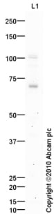 Western blot - Anti-NSMase2 antibody (ab85017)Anti-NSMase2 antibody (ab85017) at 1 µg/ml + Brain (Rat) Tissue Lysate at 10 µg
Secondary
Goat polyclonal to Rabbit IgG - H&L - Pre-Adsorbed (HRP) at 1/3000 dilution
Developed using the ECL technique.
Performed under reducing conditions.
Predicted band size: 71 kDa
Observed band size: 71 kDa
Additional bands at: 102 kDa. We are unsure as to the identity of these extra bands.
Exposure time: 20 minutes -
 Immunocytochemistry/ Immunofluorescence - Anti-NSMase2 antibody (ab85017)
ICC/IF image of ab85017 stained B35 cells. The cells were fixed with 100% methanol (5 min), permeabilized with 0.1% Triton X-100 for 5 minutes and then blocked with 1% BSA/10% normal goat serum/0.3M glycine in 0.1%PBS-Tween for 1h. The cells were then incubated with the antibody (ab85017, 5µg/ml) overnight at +4°C and ab195887, Mouse monoclonal to alpha Tubulin (Alexa Fluor® 488), at a 1/250 dilution (shown in green). The secondary antibody (shown in red) was Alexa Fluor® 647 goat anti-rabbit IgG (H+L) ab150083 used at a 1/1000 dilution for 1h. Nuclear DNA was labelled with DAPI (shown in blue).
Image was taken with a confocal microscope (Leica-Microsystems, TCS SP8).
-
 Immunocytochemistry/ Immunofluorescence - Anti-NSMase2 antibody (ab85017)ICC/IF image of ab85017 stained PC12 cells. The cells were 4% PFA fixed (10 min) and then incubated in 1%BSA / 10% normal goat serum / 0.3M glycine in 0.1% PBS-Tween for 1h to permeabilise the cells and block non-specific protein-protein interactions. The cells were then incubated with the antibody (ab85017, 5µg/ml) overnight at +4°C. The secondary antibody (green) was Alexa Fluor® 488 goat anti-rabbit IgG (H+L) used at a 1/1000 dilution for 1h. Alexa Fluor® 594 WGA was used to label plasma membranes (red) at a 1/200 dilution for 1h. DAPI was used to stain the cell nuclei (blue) at a concentration of 1.43µM. This antibody also gave a positive result in 100% methanol fixed (5 min) PC12 cells at 5µg/ml.
Protocols
Datasheets and documents
-
SDS download
-
Datasheet download
References (1)
ab85017 has been referenced in 1 publication.
- Chen L et al. Pathways of production and delivery of hepatocyte exosomes. J Cell Commun Signal 12:343-357 (2018). WB ; Mouse . PubMed: 29063370