Anti-Phospholipase C gamma 1/PLC-gamma-1 (phospho Y783) antibody (ab4828)
 antibody (ab4828)")
Key features and details
- Rabbit polyclonal to Phospholipase C gamma 1/PLC-gamma-1 (phospho Y783)
- Suitable for: IHC-P, WB
- Reacts with: Mouse, Human
- Isotype: IgG
Get better batch-to-batch reproducibility with a recombinant antibody


- Research with confidence – consistent and reproducible results with every batch
- Long-term and scalable supply – powered by recombinant technology for fast production
- Success from the first experiment – confirmed specificity through extensive validation
- Ethical standards compliant – production is animal-free
Overview
-
Product name
Anti-Phospholipase C gamma 1/PLC-gamma-1 (phospho Y783) antibody
See all Phospholipase C gamma 1/PLC-gamma-1 primary antibodies -
Description
Rabbit polyclonal to Phospholipase C gamma 1/PLC-gamma-1 (phospho Y783) -
Host species
Rabbit -
Specificity
Phosphorylation site-specific antibody selective for the phosphorylated form of the Phospholipase C gamma 1/PLC-gamma-1 containing a phosphate on tyrosine 783. The antibody has been shown to recognize the endogenous form of Phospholipase C gamma 1/PLC-gamma-1 when phosphorylated on tyrosine 783 in a variety of cell types following treatment by a broad range of extracellular stimuli. Applications include NIH 3T3 cells +/- PDGF. The antibody does not react with a site directed mutant (Y783F).
-
Tested applications
Suitable for: IHC-P, WBmore details -
Species reactivity
Reacts with: Mouse, Human
Predicted to work with: Rat, Cow, a wide range of other species
-
Immunogen
Synthetic peptide corresponding to Human Phospholipase C gamma 1/PLC-gamma-1 (phospho Y783). This region is conserved among many species including mouse, rat, cow and frog.
-
General notes
The Life Science industry has been in the grips of a reproducibility crisis for a number of years. Abcam is leading the way in addressing this with our range of recombinant monoclonal antibodies and knockout edited cell lines for gold-standard validation. Please check that this product meets your needs before purchasing.
If you have any questions, special requirements or concerns, please send us an inquiry and/or contact our Support team ahead of purchase. Recommended alternatives for this product can be found below, along with publications, customer reviews and Q&As
Properties
-
Form
Liquid -
Storage instructions
Shipped at 4°C. Upon delivery aliquot and store at -20°C or -80°C. Avoid repeated freeze / thaw cycles. -
Storage buffer
pH: 7.30
Preservative: 0.05% Sodium azide
Constituents: PBS, 50% Glycerol, 0.1% BSA
BSA is IgG and protease free -
 Concentration information loading...
 -
Purity
Immunogen affinity purified -
Purification notes
Purified from rabbit serum by sequential epitope-specific chromatography. The antibody has been negatively preadsorbed using i) a non-phosphopeptide corresponding to the site of phosphorylation to remove antibody that is reactive with nonphosphorylated Phospholipase C gamma 1/PLC-gamma-1 enzymes and (ii) a generic tyrosine phosphorylated peptide to remove antibody that is reactive with phosphotyrosine, irrespective of the sequence. The final product is generated by affinity chromatography using an Phospholipase C gamma 1/PLC-gamma-1 derived peptide that is phosphorylated at tyrosine 783. -
Clonality
Polyclonal -
Isotype
IgG -
Research areas
Associated products
-
Compatible Secondaries
-
Isotype control
-
Recombinant Protein
Applications
The Abpromise guarantee
Our Abpromise guarantee covers the use of ab4828 in the following tested applications.
The application notes include recommended starting dilutions; optimal dilutions/concentrations should be determined by the end user.
| Application | Abreviews | Notes |
|---|---|---|
| IHC-P |
Use at an assay dependent concentration.
|
|
| WB | (1) |
1/1000. Predicted molecular weight: 135 kDa.
|
| Notes |
|---|
|
IHC-P
Use at an assay dependent concentration. |
|
WB
1/1000. Predicted molecular weight: 135 kDa. |
Target
-
Function
Plays a role in actin reorganization and cell migration. The production of the second messenger molecules diacylglycerol (DAG) and inositol 1,4,5-trisphosphate (IP3) is mediated by activated phosphatidylinositol-specific phospholipase C enzymes. Major substrate for heparin-binding growth factor 1 (acidic fibroblast growth factor)-activated tyrosine kinase. -
Sequence similarities
Contains 1 C2 domain.
Contains 1 EF-hand domain.
Contains 2 PH domains.
Contains 1 PI-PLC X-box domain.
Contains 1 PI-PLC Y-box domain.
Contains 2 SH2 domains.
Contains 1 SH3 domain. -
Domain
The SH3 domain mediates interaction with CLNK (By similarity). The SH3 domain also mediates interaction with RALGPS1. -
Post-translational
modificationsThe receptor-mediated activation of PLC-gamma-1 and PLC-gamma-2 involves their phosphorylation by tyrosine kinases in response to ligation of a variety of growth factor receptors and immune system receptors. May be dephosphorylated by PTPRJ.
Ubiquitinated by CBLB in activated T-cells. -
Cellular localization
Cell projection > lamellipodium. Cell projection > ruffle. Rapidly redistributed to ruffles and lamellipodia structures in response to epidermal growth factor (EGF) treatment. - Information by UniProt
-
Database links
- Entrez Gene: 281987 Cow
- Entrez Gene: 5335 Human
- Entrez Gene: 18803 Mouse
- Entrez Gene: 25738 Rat
- Omim: 172420 Human
- SwissProt: P08487 Cow
- SwissProt: P19174 Human
- SwissProt: Q62077 Mouse
see all -
Alternative names
- 1 phosphatidyl D myo inositol 4 5 bisphosphate antibody
- 1 phosphatidylinositol 4 5 bisphosphate phosphodiesterase gamma 1 antibody
- 1-phosphatidylinositol-4,5-bisphosphate phosphodiesterase gamma-1 antibody
see all
Images
-
Western blot - Anti-Phospholipase C gamma 1/PLC-gamma-1 (phospho Y783) antibody (ab4828)
Extracts prepared from NIH/3T3 (Mouse embryonic fibroblast cell line) cells exposed to PDGF (10 ng/mL) for 10 minutes were resolved on a 10% Tris-glycine gel and transferred to nitrocellulose. Membranes were blocked with 1% BSA, followed by incubation with 0.5 µg/mL ab4828 antibody. After washing, membranes were incubated with goat F(ab')2 anti-rabbit IgG alkaline phosphatase and the signal was detected by chemiluminescence using the Tropix WesternStar detection method and Kodak BioMax ultraclear film. The data show that PDGF is a strong inducer of Phospholipase C gamma 1/PLC-gamma-1 phosphorylation on Y783 in NIH3T3 cells. Extracts prepared from NIH-3T3 cells exposed to PDGF (10 ng/mL) for 10 minutes were resolved on a 10% Tris-glycine gel and transferred to nitrocellulose. Membranes were blocked with 1% BSA, followed by incubation with 0.5 µg/mL ab4828 antibody. After washing, membranes were incubated with goat F(ab')2 anti-rabbit IgG alkaline phosphatase and the signal was detected by chemiluminescence using the Tropix
-
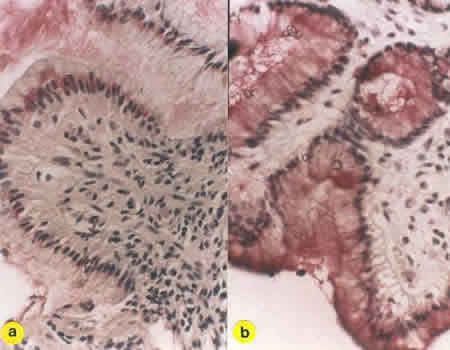 Immunohistochemistry (Formalin/PFA-fixed paraffin-embedded sections) - Anti-Phospholipase C gamma 1/PLC-gamma-1 (phospho Y783) antibody (ab4828)
ab5163 at a diluton of 1/1000 staining Phospholipase C gamma 1/PLC-gamma-1 (phospho Y783) in formalin fixed paraffin embedded sections of a) inflammed human gastric epithelium and b) normal human gastric epithelium by immunohistochemistry.
Protocols
Datasheets and documents
-
SDS download
-
Datasheet download
References (1)
ab4828 has been referenced in 1 publication.
- Bates RC et al. Activation of Src and release of intracellular calcium by phosphatidic acid during Xenopus laevis fertilization. Dev Biol N/A:N/A (2013). Xenopus laevis . PubMed: 24269904