Human BSG (CD147) knockout A549 cell line (ab273748)
 knockout A549 cell line (ab273748)")
Overview
-
Product name
Human BSG (CD147) knockout A549 cell line
See all CD147 lysates -
Parental Cell Line
A549 -
Organism
Human -
Mutation description
Knockout achieved by using CRISPR/Cas9, Homozygous: 78 bp insertion in exon 5 introducing premature STOP codon -
Passage number
<20 -
Knockout validation
Sanger Sequencing, Western Blot (WB) -
Tested applications
Suitable for: WB, Flow Cyt, Sandwich ELISAmore details -
Biosafety level
1 -
General notes
Recommended control: Human wild-type A549 cell line (ab275463). Please note a wild-type cell line is not automatically included with a knockout cell line order, if required please add recommended wild-type cell line at no additional cost using the code WILDTYPE-TMTK1.
Cryopreservation cell medium: Cell Freezing Medium-DMSO Serum free media, contains 8.7% DMSO in MEM supplemented with methyl cellulose.
Culture medium: F-12K + 10% FBS
Initial handling guidelines: Upon arrival, the vial should be stored in liquid nitrogen vapor phase and not at -80°C. Storage at -80°C may result in loss of viability.
1. Thaw the vial in 37°C water bath for approximately 1-2 minutes.
2. Transfer the cell suspension (0.8 mL) to a 15 mL/50 mL conical sterile polypropylene centrifuge tube containing 8.4 mL pre-warmed culture medium, wash vial with an additional 0.8 mL culture medium (total volume 10 mL) to collect remaining cells, and centrifuge at 201 x g (rcf) for 5 minutes at room temperature. 10 mL represents minimum recommended dilution. 20 mL represents maximum recommended dilution.
3. Resuspend the cell pellet in 5 mL pre-warmed culture medium and count using a haemocytometer or alternative cell counting method. Based on cell count, seed cells in an appropriate cell culture flask at a density of 2x10³-1x10⁴ cells/cm2. Seeding density is given as a guide only and should be scaled to align with individual lab schedules.
4. Incubate the culture at 37°C incubator with 5% CO2. Cultures should be monitored daily.Subculture guidelines:
- All seeding densities should be based on cell counts gained by established methods.
- A guide seeding density of 6x10⁴ cells/cm2 is recommended.
- A partial media change 24 hours prior to subculture may be helpful to encourage growth, if required.
- Cells should be passaged when they have achieved 80-90% confluence.
- Do not exceed 7x10⁴ cells/cm2.
This product is subject to limited use licenses from The Broad Institute, ERS Genomics Limited and Sigma-Aldrich Co. LLC, and is developed with patented technology. For full details of the licenses and patents please refer to our limited use license and patent pages.
We will provide viable cells that proliferate on revival.
Properties
-
Number of cells
1 x 10⁶ cells/vial, 1 mL -
Adherent /Suspension
Adherent -
Tissue
Lung -
Cell type
epithelial -
Disease
Carcinoma -
Gender
Male -
Mycoplasma free
Yes -
Storage instructions
Shipped on Dry Ice. Store in liquid nitrogen. -
Storage buffer
Constituents: 8.7% Dimethylsulfoxide, 2% Cellulose, methyl ether -
Research areas
Target
-
Function
Plays pivotal roles in spermatogenesis, embryo implantation, neural network formation and tumor progression. Stimulates adjacent fibroblasts to produce matrix metalloproteinases (MMPS). May target monocarboxylate transporters SLC16A1, SLC16A3 and SLC16A8 to plasma membranes of retinal pigment epithelium and neural retina. Seems to be a receptor for oligomannosidic glycans. In vitro, promotes outgrowth of astrocytic processes. -
Tissue specificity
Present only in vascular endothelium in non-neoplastic regions of the brain, whereas it is present in tumor cells but not in proliferating blood vessels in malignant gliomas. -
Sequence similarities
Contains 1 Ig-like C2-type (immunoglobulin-like) domain.
Contains 1 Ig-like V-type (immunoglobulin-like) domain. -
Post-translational
modificationsN-glycosylated. -
Cellular localization
Cell membrane. Melanosome. Colocalizes with SLC16A1 and SLC16A8 (By similarity). Identified by mass spectrometry in melanosome fractions from stage I to stage IV. - Information by UniProt
Associated products
-
KO cell lysates
-
Related Products
Applications
The Abpromise guarantee
Our Abpromise guarantee covers the use of ab273748 in the following tested applications.
The application notes include recommended starting dilutions; optimal dilutions/concentrations should be determined by the end user.
| Application | Abreviews | Notes |
|---|---|---|
| WB |
Use at an assay dependent concentration. Predicted molecular weight: 42 kDa.
|
|
| Flow Cyt |
Use at an assay dependent concentration.
|
|
| Sandwich ELISA |
Use at an assay dependent concentration.
|
| Notes |
|---|
|
WB
Use at an assay dependent concentration. Predicted molecular weight: 42 kDa. |
|
Flow Cyt
Use at an assay dependent concentration. |
|
Sandwich ELISA
Use at an assay dependent concentration. |
Images
-
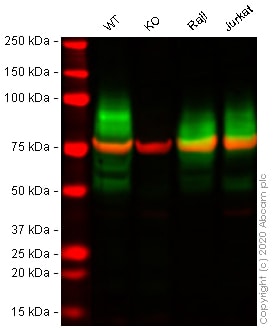
Western blot - Human BSG (CD147) knockout A549 cell line (ab273748)All lanes : Anti-CD147 antibody [MEM-M6/1] (ab666) at 1 µg/ml
Lane 1 : Wild-type A549 cell lysate
Lane 2 : BSG knockout A549 cell lysate
Lane 3 : Raji cell lysate
Lane 4 : Jurkat cell lysate
Lysates/proteins at 30 µg per lane.
Performed under reducing conditions.
Predicted band size: 42 kDa
Observed band size: 55-70 kDa why is the actual band size different from the predicted?Lanes 1 - 4: Merged signal (red and green). Green - ab666 observed at 55-70 kDa. Red - loading control ab52866 (Rabbit anti-alpha Tubulin antibody [EP1332Y]) observed at 55kDa.
ab666 was shown to react with CD147 in wild-type A549 cells in western blot with loss of signal observed in BSG knockout cell line ab273748 (knockout cell lysate ab275500). Wild-type and BSG knockout A549 cell lysates were subjected to SDS-PAGE. Membranes were blocked in fluorescent western blot (TBS-based) blocking solution before incubation with ab666 and ab52866 (Rabbit anti-alpha Tubulin antibody [EP1332Y]) overnight at 4°C at 1 µg/ml and a 1 in 20000 dilution respectively. Blots were incubated with Goat anti-Mouse IgG H&L (IRDye® 800CW) preabsorbed (ab216772) and Goat anti-Rabbit IgG H&L (IRDye® 680RD) preabsorbed (ab216777) secondary antibodies at 1 in 20000 dilution for 1 hour at room temperature before imaging.
-
 Flow Cytometry (Intracellular) - Human BSG (CD147) knockout A549 cell line (ab273748)
Flow cytometry overlay histogram showing wild-type A549 (green line) and BSG knockout A549 cells (ab273748) stained with ab666 (red line). The cells were incubated in 1x PBS containing 10% normal goat serum to block non-specific protein-protein interaction followed by the antibody (ab666) (1x10⁶ in 100μl at 10 μg/ml) for 30 min at 4°C.
The secondary antibody Goat anti-mouse IgG H&L (Alexa Fluor® 488, pre-adsorbed) (ab150117) was used at 1/2000 for 30 min at 4°C.
Isotype control antibody was mouse IgG1κ (ab170190) used at the same concentration and conditions as the primary antibody (wild-type A549 - black line; BSG knockout A549 - grey line). Unlabelled sample was also used as a control (this line is not shown for the purpose of simplicity).
Acquisition of >5000 events were collected using a 50 mW Blue laser (488nm) and 525/40 bandpass filter.
-
 Sandwich ELISA - Human BSG (CD147) knockout A549 cell line (ab273748)Human CD147 concentration was interpolated from the EMMPRIN (CD147) standard curve. Supernatants from cell culture samples were serially diluted and assessed by the Human EMMPRIN ELISA kit (ab219631). Wild-type and CD147 knockout A549 cells (ab273748) were assessed in duplicate (n=2); Jurkat and Raji cells were used as positive and negative controls respectively (n=1). Where samples were run in duplicate, data are represented as the mean and error bars represent standard deviation.
 -
 Western blot - Human BSG (CD147) knockout A549 cell line (ab273748)All lanes : Anti-CD147 antibody [EPR4053] (ab108308) at 1/1000 dilution
Lane 1 : Wild-type A549 cell lysate
Lane 2 : BSG knockout A549 cell lysate
Lane 3 : Raji cell lysate
Lysates/proteins at 30 µg per lane.
Performed under reducing conditions.
Predicted band size: 42 kDa
Observed band size: 42-70 kDa why is the actual band size different from the predicted?Lanes 1 - 3: Merged signal (red and green). Green - ab108308 observed at 42-70 kDa. Red - loading control ab7291 (Mouse anti-Alpha Tubulin [DM1A] observed at 55kDa.
ab108308 was shown to react with CD147 in wild-type A549 cells in western blot with loss of signal observed in BSG knockout cell line ab273748 (knockout cell lysate ab275500). Wild-type and BSG knockout A549 cell lysates were subjected to SDS-PAGE. Membranes were blocked in fluorescent western blot (TBS-based) blocking solution before incubation with ab108308 and ab7291 (Mouse anti-Alpha Tubulin [DM1A] overnight at 4°C at a 1 in 1000 Dilution and a 1 in 20000 dilution respectively. Blots were incubated with Goat anti-Rabbit IgG H&L (IRDye® 800CW) preabsorbed (ab216773) and Goat anti-Mouse IgG H&L (IRDye® 680RD) preabsorbed (ab216776) secondary antibodies at 1 in 20000 dilution for 1 hour at room temperature before imaging.
-
 Western blot - Human BSG (CD147) knockout A549 cell line (ab273748)All lanes : Anti-CD147 antibody [OTI9B10] (ab119020) at 1/2000 dilution
Lane 1 : Wild-type A549 cell lysate
Lane 2 : BSG knockout A549 cell lysate
Lane 3 : Raji cell lysate
Lane 4 : Jurkat cell lysate
Lysates/proteins at 30 µg per lane.
Performed under reducing conditions.
Predicted band size: 42 kDa
Observed band size: 55-70 kDa why is the actual band size different from the predicted?Lanes 1 - 4: Merged signal (red and green). Green - ab119020 observed at 55-70 kDa. Red - loading control ab52866 (Rabbit anti-alpha Tubulin antibody [EP1332Y]) observed at 55kDa.
ab119020 was shown to react with CD147 in wild-type A549 cells in western blot with loss of signal observed in BSG knockout cell line ab273748 (knockout cell lysate ab275500). Wild-type and BSG knockout A549 cell lysates were subjected to SDS-PAGE. Membranes were blocked in fluorescent western blot (TBS-based) blocking solution before incubation with ab119020 and ab52866 (Rabbit anti-alpha Tubulin antibody [EP1332Y]) overnight at 4°C at a 1 in 2000 Dilution and a 1 in 20000 dilution respectively. Blots were incubated with Goat anti-Mouse IgG H&L (IRDye® 800CW) preabsorbed (ab216772) and Goat anti-Rabbit IgG H&L (IRDye® 680RD) preabsorbed (ab216777) secondary antibodies at 1 in 20000 dilution for 1 hour at room temperature before imaging.
-
 Western blot - Human BSG (CD147) knockout A549 cell line (ab273748)All lanes : Anti-CD147 antibody [10E10] (ab230921) at 1/500 dilution
Lane 1 : Wild-type A549 cell lysate
Lane 2 : BSG knockout A549 cell lysate
Lane 3 : Raji cell lysate
Lane 4 : Jurkat cell lysate
Lysates/proteins at 30 µg per lane.
Performed under reducing conditions.
Predicted band size: 42 kDa
Observed band size: 42-70 kDa why is the actual band size different from the predicted?Lanes 1 - 4: Merged signal (red and green). Green - ab230921 observed at 42-70 kDa. Red - loading control ab52866 (Rabbit anti-alpha Tubulin antibody [EP1332Y]) observed at 55kDa.
ab230921 was shown to react with CD147 in wild-type A549 cells in western blot with loss of signal observed in BSG knockout cell line ab273748 (knockout cell lysate ab275500). Wild-type and BSG knockout A549 cell lysates were subjected to SDS-PAGE. Membranes were blocked in fluorescent western blot (TBS-based) blocking solution before incubation with ab230921 and ab52866 (Rabbit anti-alpha Tubulin antibody [EP1332Y]) overnight at 4°C at a 1 in 500 Dilution and a 1 in 20000 dilution respectively. Blots were incubated with Goat anti-Mouse IgG H&L (IRDye® 800CW) preabsorbed (ab216772) and Goat anti-Rabbit IgG H&L (IRDye® 680RD) preabsorbed (ab216777) secondary antibodies at 1 in 20000 dilution for 1 hour at room temperature before imaging.
-
 Flow Cytometry - Human BSG (CD147) knockout A549 cell line (ab273748)
Flow cytometry overlay histogram showing wild-type A549 (green line) and BSG knockout A549 cells (ab273748) stained with ab194401 (red line). The cells were incubated in 1x PBS containing 10% normal goat serum to block non-specific protein-protein interaction followed by the antibody (ab194401) (1x10⁶ in 100μl at 10 μg/ml) for 30 min at 4°C.
The secondary antibody Goat anti-mouse IgG H&L (Alexa Fluor® 488, pre-adsorbed) (ab150117) was used at 1/2000 for 30 min at 4°C.
Isotype control antibody was mouse IgG1κ (ab170190) used at the same concentration and conditions as the primary antibody (wild-type A549 - black line; BSG knockout A549 - grey line). Unlabelled sample was also used as a control (this line is not shown for the purpose of simplicity).
Acquisition of >5000 events were collected using a 50 mW Blue laser (488nm) and 525/40 bandpass filter.
-
 Flow Cytometry - Human BSG (CD147) knockout A549 cell line (ab273748)
Flow cytometry overlay histogram showing wild-type A549 (green line) and BSG knockout A549 cells (ab273748) stained with ab91147 (red line). The cells were incubated in 1x PBS containing 10% normal goat serum to block non-specific protein-protein interaction followed by the antibody (ab91147) (1x10⁶ in 100μl at 10 μg/ml) for 30 min at 4°C.
The secondary antibody Goat anti-mouse IgG H&L (Alexa Fluor® 488, pre-adsorbed) (ab150117) was used at 1/2000 for 30 min at 4°C.
Isotype control antibody was mouse IgG1κ (ab170190) used at the same concentration and conditions as the primary antibody (wild-type A549 - black line; BSG knockout A549 - grey line). Unlabelled sample was also used as a control (this line is not shown for the purpose of simplicity).
Acquisition of >5000 events were collected using a 50 mW Blue laser (488nm) and 525/40 bandpass filter.
-
 Flow Cytometry - Human BSG (CD147) knockout A549 cell line (ab273748)
Flow cytometry overlay histogram showing wild-type A549 (green line) and BSG knockout A549 cells (ab273748) stained with ab21903 (red line). The cells were incubated in 1x PBS containing 10% normal goat serum to block non-specific protein-protein interaction followed by the antibody (ab21903) (1x10⁶ in 100μl at 5 μg/ml) for 30 min at 4°C.
The secondary antibody Goat anti-mouse IgG H&L (Alexa Fluor® 488, pre-adsorbed) (ab150117) was used at 1/2000 for 30 min at 4°C.
Isotype control antibody was mouse IgG2bκ (ab170192) used at the same concentration and conditions as the primary antibody (wild-type A549 - black line; BSG knockout A549 - grey line). Unlabelled sample was also used as a control (this line is not shown for the purpose of simplicity).
Acquisition of >5000 events were collected using a 50 mW Blue laser (488nm) and 525/40 bandpass filter.
-
 Flow Cytometry - Human BSG (CD147) knockout A549 cell line (ab273748)
Flow cytometry overlay histogram showing wild-type A549 (green line) and BSG knockout A549 cells (ab273748) stained with ab230921 (red line). The cells were incubated in 1x PBS containing 10% normal goat serum to block non-specific protein-protein interaction followed by the antibody (ab230921) (1x10⁶ in 100μl at 5 μg/ml) for 30 min at 4°C.
The secondary antibody Goat anti-mouse IgG H&L (Alexa Fluor® 488, pre-adsorbed) (ab150117) was used at 1/2000 for 30 min at 4°C.
Isotype control antibody was mouse IgG1κ (ab170190) used at the same concentration and conditions as the primary antibody (wild-type A549 - black line; BSG knockout A549 - grey line). Unlabelled sample was also used as a control (this line is not shown for the purpose of simplicity).
Acquisition of >5000 events were collected using a 50 mW Blue laser (488nm) and 525/40 bandpass filter.
-
 Sanger Sequencing - Human BSG (CD147) knockout A549 cell line (ab273748)
Allele-1: 78 bp insertion in exon 5 introducing premature STOP codon.
Protocols
Datasheets and documents
-
SDS download
-
Datasheet download
References (0)
ab273748 has not yet been referenced specifically in any publications.