Recombinant AMARV GP protein (His tag) (ab190126)
 (ab190126)")
Key features and details
- Expression system: Baculovirus infected Sf9 cells
- Purity: > 70% SDS-PAGE
- Tags: His tag C-Terminus
- Suitable for: WB, SDS-PAGE, ELISA
Description
-
Product name
Recombinant AMARV GP protein (His tag) -
Purity
> 70 % SDS-PAGE. -
Expression system
Baculovirus infected Sf9 cells -
Accession
-
Protein length
Protein fragment -
Animal free
No -
Nature
Recombinant -
-
Species
Marburg virus -
Sequence
MKTTCLLISLILIQGVKTLPILEIASNIQPQNVDSVCSGTLQKTEDVHLM GFTLSGQKVADSPLEASKRW AFRAGVPPKNVEYTEGEEAKTCYNISVT DPSGKSLLLDPPTNIRDYPKCKTIHHIQGQNPHAQGIALHLW GAFFLY DRIASTTMYRGKVFTEGNIAAMIVNKTVHKMIFSRQGQGYRHMNLTSTNK YWTSSNGTQTNDTG CFGTLQEYNSTKNQTCAPSKKPLPLPTAHPEVKL TSTSTDATKLNTTDPNSDDEDLTTSGSGSGEQEPYT TSDAATKQGLSS TMPPTPSPQPSTPQQGGNNTNHSQGVVTEPGKTNTTAQPSMPPHNTTTIS TNNTSKHN LSTPSVPIQNATNYNTQSTAPENEQTSAPSKTTLLPTENP TTAKSTNSTKSPTTTVPNTTNKYSTSPSPT PDSTAQHLVYFRRKRNIL WREGDMFPFLDGLINAPIDFDPVPNTKTIFDESSSSGASAEEDQHASPNI SL TLSYFPKVNENTAHSGENENDCDAELRIWSVQEDDLAAGLSWIPFF GPGIEGLYTAGLIKNQNNLVCRLR RLANQTAKSLELLLRVTTEERTFS LINRHAIDFLLARWGGTCKVLGPDCCIGIEDLSRNISEQIDQIKKD EQ KEGTGWGLGGKWWTSDWGVLTNLGILLLLSIAVLIALSCICRIFTKYIG -
Predicted molecular weight
60 kDa including tags -
Tags
His tag C-Terminus -
Additional sequence information
Angola marburgvirus glycoprotein minus the transmembrane domain (MARV-Angola rGPdTM). The theoretical molecular weight of the protein is ~60 kDa including the His-tag, without glycosylation.
-
Associated products
-
Related Products
Specifications
Our Abpromise guarantee covers the use of ab190126 in the following tested applications.
The application notes include recommended starting dilutions; optimal dilutions/concentrations should be determined by the end user.
-
Applications
Western blot
SDS-PAGE
ELISA
-
Form
Liquid -
 Concentration information loading...
Preparation and Storage
-
Stability and Storage
Shipped at 4°C. Store at -80°C. Avoid freeze / thaw cycle.
Constituent: PBS
PBS is supplemented with 10% glycerol, arginine and glutamic acid.
General Info
-
Alternative names
- Envelope glycoprotein
- GP
- GP1
see all -
Relevance
GP1 is responsible for binding to the receptor(s) on target cells. Interacts with CD209/DC-SIGN and CLEC4M/DC-SIGNR which act as cofactors for virus entry into the host cell. Binding to CD209 and CLEC4M, which are respectively found on dendritic cells (DCs), and on endothelial cells of liver sinusoids and lymph node sinuses, facilitate infection of macrophages and endothelial cells. These interactions not only facilitate virus cell entry, but also allow capture of viral particles by DCs and subsequent transmission to susceptible cells without DCs infection (trans infection). GP2 acts as a class I viral fusion protein. Under the current model, the protein has at least 3 conformational states: pre-fusion native state, pre-hairpin intermediate state, and post-fusion hairpin state. During viral and target cell membrane fusion, the coiled coil regions (heptad repeats) assume a trimer-of-hairpins structure, positioning the fusion peptide in close proximity to the C-terminal region of the ectodomain. The formation of this structure appears to drive apposition and subsequent fusion of viral and target cell membranes. Responsible for penetration of the virus into the cell cytoplasm by mediating the fusion of the membrane of the endocytosed virus particle with the endosomal membrane. Low pH in endosomes induces an irreversible conformational change in GP2, releasing the fusion hydrophobic peptide -
Cellular localization
GP2: Virion membrane; Single-pass type I membrane protein. Virion membrane; Lipid-anchor. Host cell membrane; Single-pass type I membrane protein . Host cell membrane; Lipid-anchor By similarity. Note: In the cell, localizes to the plasma membrane lipid rafts, which probably represent the assembly and budding site. GP1: Virion membrane; Peripheral membrane protein. Host cell membrane; Peripheral membrane protein. Note: GP1 is not anchored to the viral envelope, but associates with the extravirion surface through its binding to GP2. In the cell, both GP1 and GP2 localize to the plasma membrane lipid rafts, which probably represent the assembly and budding site.
Images
-
Western blot - Recombinant AMARV GP protein (His tag) (ab190126)All lanes : rabbit polyclonal antibody anti AMARV GP at 0.5 µg/ml
Lane 1 :Recombinant AMARV GP protein (His tag) (ab190126) at 0.1 µg
Lane 2 :Recombinant AMARV GP protein (His tag) (ab190126) at 0.5 µg
Lane 3 :Recombinant AMARV GP protein (His tag) (ab190126) at 1 µg
Secondary
All lanes : anti-rabbit IgG-HRP conjugate, followed by substrate.
Predicted band size: 74 kDa -
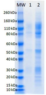 SDS-PAGE - Recombinant AMARV GP protein (His tag) (ab190126)
SDS-PAGE analysis of 1 μg and 5 μg (lanes 1, 2 respectively) of ab190126 under denaturing and reducing conditions.
-
 ELISA - Recombinant AMARV GP protein (His tag) (ab190126)
Plate was coated with ab190126 starting at 800 ng/well, serially diluted in DPBS. Washed plate was detected using one dilution of a positive control serum, followed with anti-IgG HRP conjugate and TM substrate. OD650 is reported. Background of ab190126 coated plate without positive control serum was 0.051 OD650.
Protocols
To our knowledge, customised protocols are not required for this product. Please try the standard protocols listed below and let us know how you get on.
Datasheets and documents
-
Datasheet download
References (0)
ab190126 has not yet been referenced specifically in any publications.