Anti-Synaptophysin antibody (ab32594)
")
Key features and details
- Rabbit polyclonal to Synaptophysin
- Suitable for: WB, ICC/IF
- Reacts with: Rat
- Isotype: IgG
Get better batch-to-batch reproducibility with a recombinant antibody


- Research with confidence – consistent and reproducible results with every batch
- Long-term and scalable supply – powered by recombinant technology for fast production
- Success from the first experiment – confirmed specificity through extensive validation
- Ethical standards compliant – production is animal-free
Overview
-
Product name
Anti-Synaptophysin antibody
See all Synaptophysin primary antibodies -
Description
Rabbit polyclonal to Synaptophysin -
Host species
Rabbit -
Tested applications
Suitable for: WB, ICC/IFmore details -
Species reactivity
Reacts with: Rat
Predicted to work with: Human
-
Immunogen
Synthetic peptide:
GPGGYGPQDSYGPQGGYQPD
, corresponding to amino acids 253-272 of Rat Synaptophysin -
Positive control
- WB: Mouse and rat brain tissue lysates. ICC/IF: rat hippocampal neurons
-
General notes
The Life Science industry has been in the grips of a reproducibility crisis for a number of years. Abcam is leading the way in addressing this with our range of recombinant monoclonal antibodies and knockout edited cell lines for gold-standard validation. Please check that this product meets your needs before purchasing.
If you have any questions, special requirements or concerns, please send us an inquiry and/or contact our Support team ahead of purchase. Recommended alternatives for this product can be found below, along with publications, customer reviews and Q&As
Properties
-
Form
Liquid -
Storage instructions
Shipped at 4°C. Upon delivery aliquot and store at -20°C or -80°C. Avoid repeated freeze / thaw cycles. -
Storage buffer
Preservative: 0.05% Sodium azide
Constituents: PBS, 0.1% BSA -
 Concentration information loading...
 -
Purity
Immunogen affinity purified -
Clonality
Polyclonal -
Isotype
IgG -
Research areas
Associated products
-
Compatible Secondaries
-
Isotype control
-
Positive Controls
-
Recombinant Protein
Applications
The Abpromise guarantee
Our Abpromise guarantee covers the use of ab32594 in the following tested applications.
The application notes include recommended starting dilutions; optimal dilutions/concentrations should be determined by the end user.
| Application | Abreviews | Notes |
|---|---|---|
| WB | (5) |
1/5000. Detects a band of approximately 33.8 kDa (predicted molecular weight: 34 kDa).
|
| ICC/IF | (1) |
Use at an assay dependent concentration.
|
| Notes |
|---|
|
WB
1/5000. Detects a band of approximately 33.8 kDa (predicted molecular weight: 34 kDa). |
|
ICC/IF
Use at an assay dependent concentration. |
Target
-
Function
Possibly involved in structural functions as organizing other membrane components or in targeting the vesicles to the plasma membrane. Involved in the regulation of short-term and long-term synaptic plasticity. -
Tissue specificity
Characteristic of a type of small (30-80 nm) neurosecretory vesicles, including presynaptic vesicles, but also vesicles of various neuroendocrine cells of both neuronal and epithelial phenotype. -
Involvement in disease
Mental retardation, X-linked, SYP-related -
Sequence similarities
Belongs to the synaptophysin/synaptobrevin family.
Contains 1 MARVEL domain. -
Domain
The calcium-binding activity is thought to be localized in the cytoplasmic tail of the protein. -
Post-translational
modificationsUbiquitinated; mediated by SIAH1 or SIAH2 and leading to its subsequent proteasomal degradation. -
Cellular localization
Cytoplasmic vesicle, secretory vesicle, synaptic vesicle membrane. Cell junction, synapse, synaptosome. - Information by UniProt
-
Database links
- Entrez Gene: 6855 Human
- Entrez Gene: 24804 Rat
- Omim: 313475 Human
- SwissProt: P08247 Human
- SwissProt: P07825 Rat
- Unigene: 632804 Human
- Unigene: 11067 Rat
-
Alternative names
- Major synaptic vesicle protein p38 antibody
- MRX96 antibody
- MRXSYP antibody
see all
Images
-
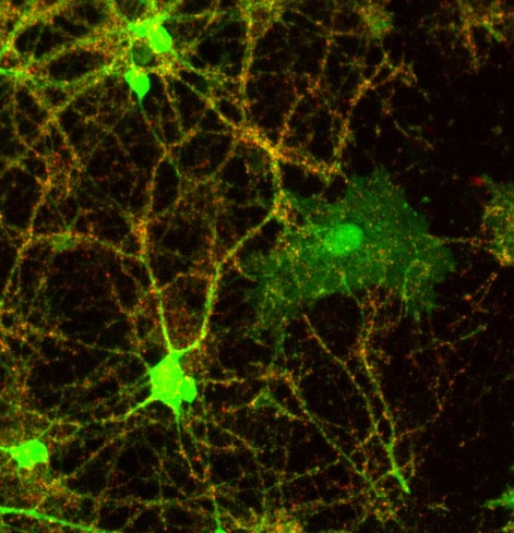
Immunocytochemistry/ Immunofluorescence - Anti-Synaptophysin antibody (ab32594)
ab32594 labelling Synaptophysin by Immunocytochemistry/Immunofluorescence in rat hippocampal neurons. The cells were fixed with 1% formaldehyde in fume hood at room temperature for 10 minutes.
ab32594 is used at 1:1500 dilution and DyLight 649 Conjugated Goat anti-Rabbit IgG used as secondary antibody at 1:1500 dilution.
-
 Western blot - Anti-Synaptophysin antibody (ab32594)All lanes : Anti-Synaptophysin antibody (ab32594) at 1/1000 dilution
Lane 1 : Mouse brain tissue lysate
Lane 2 : Rat brain tissue lysate
Lane 3 : Rat liver tissue lysate
Lane 4 : Rat kidney tissue lysate
Lysates/proteins at 30 µg per lane.
Predicted band size: 34 kDaWestern blot analysis was performed on tissue extracts (30 µg lysate). The blot was probed with ab32594 (1:1000 dilution) and detected by chemiluminescence using Goat anti-Rabbit IgG (H+L) Superclonal™ Secondary Antibody, HRP conjugate. A 34 kDa band corresponding to Synaptophysin was observed in Mouse Brain, Rat Brain and not observed in other tissues which are documented to be Synaptophysin negative.
-
 Western blot - Anti-Synaptophysin antibody (ab32594)This image is courtesy of an anonymous AbreviewAnti-Synaptophysin antibody (ab32594) at 1/1000 dilution + Apteronotus leptorhynchus brain tissue lysate at 50 µg
Secondary
AlexaFluor®488-conjugated goat anti-rabbit polyclonal IgG
at 1/1000 dilution
Developed using the ECL technique.
Performed under reducing conditions.
Predicted band size: 34 kDa
Observed band size: 33,35 kDa why is the actual band size different from the predicted?
Exposure time: 7 minutes
-
 Immunocytochemistry/ Immunofluorescence - Anti-Synaptophysin antibody (ab32594)
ab32594 labelling Synaptophysin by Immunocytochemistry/Immunofluorescence in rat hippocampal neurons. The cells were fixed with 1% formaldehyde in fume hood at room temperature for 10 minutes.
ab32594 is used at 1:1500 dilution and DyLight 649 Conjugated Goat anti-Rabbit IgG used as secondary antibody at 1:1500 dilution.
-
 Western blot - Anti-Synaptophysin antibody (ab32594)Anti-Synaptophysin antibody (ab32594) at 1/5000 dilution + Rat brain
Predicted band size: 34 kDa
Observed band size: 33.8 kDa why is the actual band size different from the predicted?
Protocols
Datasheets and documents
-
SDS download
-
Datasheet download
References (54)
ab32594 has been referenced in 54 publications.
- Kútna V et al. Cerebellar demyelination and neurodegeneration associated with mTORC1 hyperactivity may contribute to the developmental onset of autism-like neurobehavioral phenotype in a rat model. Autism Res 15:791-805 (2022). PubMed: 35178882
- Yao L et al. Extrasynaptic NMDA Receptors Bidirectionally Modulate Intrinsic Excitability of Inhibitory Neurons. J Neurosci 42:3066-3079 (2022). PubMed: 35197319
- Wang F et al. Low-Intensity Focused Ultrasound Stimulation Ameliorates Working Memory Dysfunctions in Vascular Dementia Rats via Improving Neuronal Environment. Front Aging Neurosci 14:814560 (2022). PubMed: 35264943
- Hyun SA et al. Bisphenol-A impairs synaptic formation and function by RGS4-mediated regulation of BDNF signaling in the cerebral cortex. Dis Model Mech 15:N/A (2022). PubMed: 35781563
- Olguin SL et al. KHSRP loss increases neuronal growth and synaptic transmission and alters memory consolidation through RNA stabilization. Commun Biol 5:672 (2022). PubMed: 35798971